5KJ1

G173A horse liver alcohol dehydrogenase complexed with NAD+ and pentafluorobenzyl alcohol

  • Classification: OXIDOREDUCTASE
  • Organism(s): Equus caballus
  • Expression System: Escherichia coli
  • Mutation(s): Yes 

  • Deposited: 2016-06-17 Released: 2016-07-06 
  • Deposition Author(s): Plapp, B.V.
  • Funding Organization(s): National Institutes of Health/National Institute of General Medical Sciences (NIH/NIGMS)

Experimental Data Snapshot

  • Method: X-RAY DIFFRACTION
  • Resolution: 1.20 Å
  • R-Value Free: 0.170 
  • R-Value Work: 0.124 
  • R-Value Observed: 0.124 

wwPDB Validation   3D Report Full Report


Ligand Structure Quality Assessment 


This is version 1.7 of the entry. See complete history


Literature

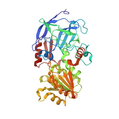
Dependence of crystallographic atomic displacement parameters on temperature (25-150 K) for complexes of horse liver alcohol dehydrogenase.

Plapp, B.V.Gakhar, L.Subramanian, R.

(2022) Acta Crystallogr D Struct Biol 78: 1221-1234

  • DOI: https://doi.org/10.1107/S2059798322008361
  • Primary Citation of Related Structures:  
    5KJ1

  • PubMed Abstract: 

    Enzymes catalyze reactions by binding and orienting substrates with dynamic interactions. Horse liver alcohol dehydrogenase catalyzes hydrogen transfer with quantum-mechanical tunneling that involves fast motions in the active site. The structures and B factors of ternary complexes of the enzyme with NAD + and 2,3,4,5,6-pentafluorobenzyl alcohol or NAD + and 2,2,2-trifluoroethanol were determined to 1.1-1.3 Å resolution below the `glassy transition' in order to extract information about the temperature-dependent harmonic motions, which are reflected in the crystallographic B factors. The refinement statistics and structures are essentially the same for each structure at all temperatures. The B factors were corrected for a small amount of radiation decay. The overall B factors for the complexes are similar (13-16 Å 2 ) over the range 25-100 K, but increase somewhat at 150 K. Applying TLS refinement to remove the contribution of pseudo-rigid-body displacements of coenzyme binding and catalytic domains provided residual B factors of 7-10 Å 2 for the overall complexes and of 5-10 Å 2 for C4N of NAD + and the methylene carbon of the alcohols. These residual B factors have a very small dependence on temperature and include local harmonic motions and apparently contributions from other sources. Structures at 100 K show complexes that are poised for hydrogen transfer, which involves atomic displacements of ∼0.3 Å and is compatible with the motions estimated from the residual B factors and molecular-dynamics simulations. At 298 K local conformational changes are also involved in catalysis, as enzymes with substitutions of amino acids in the substrate-binding site have similar positions of NAD + and pentafluorobenzyl alcohol and similar residual B factors, but differ by tenfold in the rate constants for hydride transfer.


  • Organizational Affiliation

    Department of Biochemistry and Molecular Biology, The University of Iowa, Iowa City, IA 52252, USA.


Macromolecules
Find similar proteins by:  (by identity cutoff)  |  3D Structure
Entity ID: 1
MoleculeChains Sequence LengthOrganismDetailsImage
Alcohol dehydrogenase E chain
A, B
374Equus caballusMutation(s): 1 
EC: 1.1.1.1
UniProt
Find proteins for P00327 (Equus caballus)
Explore P00327 
Go to UniProtKB:  P00327
Entity Groups  
Sequence Clusters30% Identity50% Identity70% Identity90% Identity95% Identity100% Identity
UniProt GroupP00327
Sequence Annotations
Expand
  • Reference Sequence
Small Molecules
Ligands 4 Unique
IDChains Name / Formula / InChI Key2D Diagram3D Interactions
NAJ
Query on NAJ

Download Ideal Coordinates CCD File 
E [auth A],
L [auth B]
NICOTINAMIDE-ADENINE-DINUCLEOTIDE (ACIDIC FORM)
C21 H27 N7 O14 P2
BAWFJGJZGIEFAR-NNYOXOHSSA-N
PFB
Query on PFB

Download Ideal Coordinates CCD File 
F [auth A],
M [auth B]
2,3,4,5,6-PENTAFLUOROBENZYL ALCOHOL
C7 H3 F5 O
PGJYYCIOYBZTPU-UHFFFAOYSA-N
MRD
Query on MRD

Download Ideal Coordinates CCD File 
G [auth A],
H [auth A],
I [auth A],
N [auth B]
(4R)-2-METHYLPENTANE-2,4-DIOL
C6 H14 O2
SVTBMSDMJJWYQN-RXMQYKEDSA-N
ZN
Query on ZN

Download Ideal Coordinates CCD File 
C [auth A],
D [auth A],
J [auth B],
K [auth B]
ZINC ION
Zn
PTFCDOFLOPIGGS-UHFFFAOYSA-N
Experimental Data & Validation

Experimental Data

Unit Cell:
Length ( Å )Angle ( ˚ )
a = 44.31α = 91.82
b = 51.5β = 103.05
c = 92.42γ = 110.12
Software Package:
Software NamePurpose
REFMACrefinement
d*TREKdata reduction
d*TREKdata scaling
REFMACphasing

Structure Validation

View Full Validation Report


Ligand Structure Quality Assessment 


Entry History & Funding Information

Deposition Data

  • Released Date: 2016-07-06 
  • Deposition Author(s): Plapp, B.V.

Funding OrganizationLocationGrant Number
National Institutes of Health/National Institute of General Medical Sciences (NIH/NIGMS)United StatesGM078446

Revision History  (Full details and data files)

  • Version 1.0: 2016-07-06
    Type: Initial release
  • Version 1.1: 2017-09-27
    Changes: Author supporting evidence, Derived calculations
  • Version 1.2: 2018-01-03
    Changes: Database references
  • Version 1.3: 2018-02-28
    Changes: Database references
  • Version 1.4: 2019-12-25
    Changes: Author supporting evidence
  • Version 1.5: 2022-04-27
    Changes: Database references, Derived calculations
  • Version 1.6: 2022-10-12
    Changes: Database references
  • Version 1.7: 2023-10-18
    Changes: Data collection, Refinement description