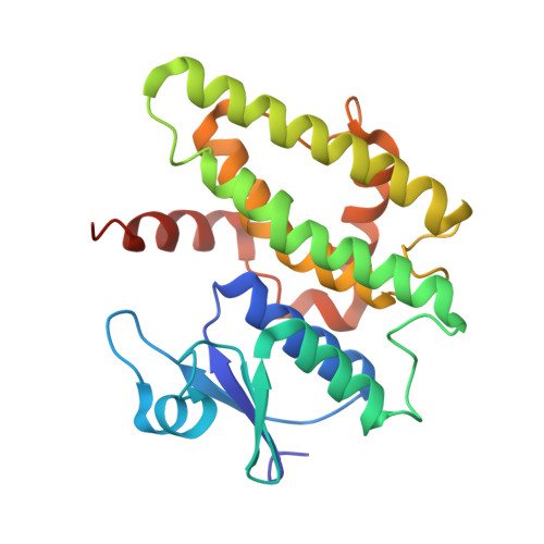
5KEJ

Crystallographic structure of the Tau class glutathione S-transferase MiGSTU in complex with S-hexyl-glutathione


Experimental Data Snapshot

  • Method: X-RAY DIFFRACTION
  • Resolution: 2.35 Å
  • R-Value Free: 0.226 
  • R-Value Work: 0.188 
  • R-Value Observed: 0.190 

wwPDB Validation   3D Report Full Report


Ligand Structure Quality Assessment 


This is version 1.2 of the entry. See complete history


Literature

Insights into ligand binding to a glutathione S-transferase from mango: Structure, thermodynamics and kinetics.

Valenzuela-Chavira, I.Contreras-Vergara, C.A.Arvizu-Flores, A.A.Serrano-Posada, H.Lopez-Zavala, A.A.Garcia-Orozco, K.D.Hernandez-Paredes, J.Rudino-Pinera, E.Stojanoff, V.Sotelo-Mundo, R.R.Islas-Osuna, M.A.

(2017) Biochimie 135: 35-45

  • DOI: https://doi.org/10.1016/j.biochi.2017.01.005
  • Primary Citation of Related Structures:  
    5G5E, 5G5F, 5KEJ

  • PubMed Abstract: 

    We studied a mango glutathione S-transferase (GST) (Mangifera indica) bound to glutathione (GSH) and S-hexyl glutathione (GSX). This GST Tau class (MiGSTU) had a molecular mass of 25.5 kDa. MiGSTU Michaelis-Menten kinetic constants were determined for their substrates obtaining a K m , V max and k cat for CDNB of 0.792 mM, 80.58 mM min -1 and 68.49 s -1 respectively and 0.693 mM, 105.32 mM min -1 and 89.57 s -1 , for reduced GSH respectively. MiGSTU had a micromolar affinity towards GSH (5.2 μM) or GSX (7.8 μM). The crystal structure of the MiGSTU in apo or bound to GSH or GSX generated a model that explains the thermodynamic signatures of binding and showed the importance of enthalpic-entropic compensation in ligand binding to Tau-class GST enzymes.


  • Organizational Affiliation

    Laboratorio de Estructura Biomolecular, Centro de Investigación en Alimentación y Desarrollo, A.C. (CIAD), Hermosillo, Sonora 83304, Mexico; Laboratorio de Genética Molecular de Plantas, Centro de Investigación en Alimentación y Desarrollo, A.C. (CIAD), Hermosillo, Sonora 83304, Mexico.


Macromolecules
Find similar proteins by:  (by identity cutoff)  |  3D Structure
Entity ID: 1
MoleculeChains Sequence LengthOrganismDetailsImage
Tau class glutathione S-transferase
A, B
229Mangifera indicaMutation(s): 0 
EC: 2.5.1.18
UniProt
Find proteins for A0A1P8NWC2 (Mangifera indica)
Explore A0A1P8NWC2 
Go to UniProtKB:  A0A1P8NWC2
Entity Groups  
Sequence Clusters30% Identity50% Identity70% Identity90% Identity95% Identity100% Identity
UniProt GroupA0A1P8NWC2
Sequence Annotations
Expand
  • Reference Sequence
Small Molecules
Binding Affinity Annotations 
IDSourceBinding Affinity
GTX Binding MOAD:  5KEJ Kd: 7800 (nM) from 1 assay(s)
Experimental Data & Validation

Experimental Data

  • Method: X-RAY DIFFRACTION
  • Resolution: 2.35 Å
  • R-Value Free: 0.226 
  • R-Value Work: 0.188 
  • R-Value Observed: 0.190 
  • Space Group: P 21 21 21
Unit Cell:
Length ( Å )Angle ( ˚ )
a = 64.319α = 90
b = 88.805β = 90
c = 96.583γ = 90
Software Package:
Software NamePurpose
XDSdata reduction
SCALAdata scaling
PHASERphasing
PHENIXrefinement

Structure Validation

View Full Validation Report


Ligand Structure Quality Assessment 


Entry History 

Deposition Data

Revision History  (Full details and data files)

  • Version 1.0: 2017-02-01
    Type: Initial release
  • Version 1.1: 2017-03-22
    Changes: Database references
  • Version 1.2: 2023-09-27
    Changes: Data collection, Database references, Refinement description