5KEI

Mycobacterium smegmatis MbtA apo structure


Experimental Data Snapshot

  • Method: X-RAY DIFFRACTION
  • Resolution: 2.33 Å
  • R-Value Free: 0.244 
  • R-Value Work: 0.189 
  • R-Value Observed: 0.192 

wwPDB Validation   3D Report Full Report


This is version 1.4 of the entry. See complete history


Literature

Post-translational Acetylation of MbtA Modulates Mycobacterial Siderophore Biosynthesis.

Vergnolle, O.Xu, H.Tufariello, J.M.Favrot, L.Malek, A.A.Jacobs, W.R.Blanchard, J.S.

(2016) J Biol Chem 291: 22315-22326

  • DOI: https://doi.org/10.1074/jbc.M116.744532
  • Primary Citation of Related Structures:  
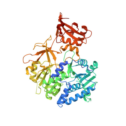
    5KEI

  • PubMed Abstract: 

    Iron is an essential element for life, but its soluble form is scarce in the environment and is rarer in the human body. Mtb (Mycobacterium tuberculosis) produces two aryl-capped siderophores, mycobactin (MBT) and carboxymycobactin (cMBT), to chelate intracellular iron. The adenylating enzyme MbtA catalyzes the first step of mycobactin biosynthesis in two half-reactions: activation of the salicylic acid as an acyl-adenylate and ligation onto the acyl carrier protein (ACP) domain of MbtB to form covalently salicylated MbtB-ACP. We report the first apo-MbtA structure from Mycobacterium smegmatis at 2.3 Å. We demonstrate here that MbtA activity can be reversibly, post-translationally regulated by acetylation. Indeed the mycobacterial Pat (protein lysine acetyltransferase), Rv0998, specifically acetylates MbtA on lysine 546, in a cAMP-dependent manner, leading to enzyme inhibition. MbtA acetylation can be reversed by the NAD + -dependent DAc (deacetyltransferase), Rv1151c. Deletion of Pat and DAc genes in Mtb revealed distinct phenotypes for strains lacking one or the other gene at low pH and limiting iron conditions. This study establishes a direct connection between the reversible acetylation system Pat/DAc and the ability of Mtb to adapt in limited iron conditions, which is critical for mycobacterial infection.


  • Organizational Affiliation

    From the Department of Biochemistry and.


Macromolecules
Find similar proteins by:  (by identity cutoff)  |  3D Structure
Entity ID: 1
MoleculeChains Sequence LengthOrganismDetailsImage
2,3-dihydroxybenzoate-AMP ligase558Mycolicibacterium smegmatis MC2 155Mutation(s): 0 
Gene Names: mbtAMSMEG_4516MSMEI_4404
EC: 6.3.2
UniProt
Find proteins for A0R0V0 (Mycolicibacterium smegmatis (strain ATCC 700084 / mc(2)155))
Explore A0R0V0 
Go to UniProtKB:  A0R0V0
Entity Groups  
Sequence Clusters30% Identity50% Identity70% Identity90% Identity95% Identity100% Identity
UniProt GroupA0R0V0
Sequence Annotations
Expand
  • Reference Sequence
Experimental Data & Validation

Experimental Data

  • Method: X-RAY DIFFRACTION
  • Resolution: 2.33 Å
  • R-Value Free: 0.244 
  • R-Value Work: 0.189 
  • R-Value Observed: 0.192 
  • Space Group: P 21 21 21
Unit Cell:
Length ( Å )Angle ( ˚ )
a = 56.362α = 90
b = 85.746β = 90
c = 104.665γ = 90
Software Package:
Software NamePurpose
PHENIXrefinement
XDSdata reduction
XDSdata scaling
PHASERphasing

Structure Validation

View Full Validation Report


Entry History & Funding Information

Deposition Data


Funding OrganizationLocationGrant Number
National Institutes of Health/National Institute Of Allergy and Infectious Diseases (NIH/NIAID)United StatesAI60899

Revision History  (Full details and data files)

  • Version 1.0: 2016-09-14
    Type: Initial release
  • Version 1.1: 2016-10-26
    Changes: Database references
  • Version 1.2: 2017-09-27
    Changes: Author supporting evidence, Database references, Derived calculations
  • Version 1.3: 2019-12-11
    Changes: Author supporting evidence
  • Version 1.4: 2023-09-27
    Changes: Data collection, Database references, Refinement description