5KDI

How FAPP2 Selects Simple Glycosphingolipids Using the GLTP-fold


Experimental Data Snapshot

  • Method: X-RAY DIFFRACTION
  • Resolution: 1.45 Å
  • R-Value Free: 0.177 
  • R-Value Work: 0.131 
  • R-Value Observed: 0.133 

wwPDB Validation   3D Report Full Report


Ligand Structure Quality Assessment 


This is version 1.3 of the entry. See complete history


Literature

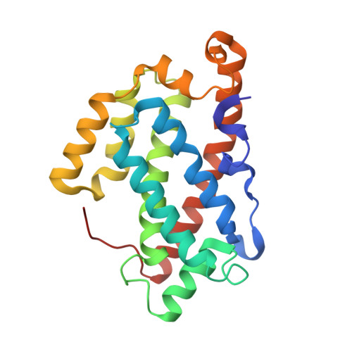
Structural analyses of 4-phosphate adaptor protein 2 yield mechanistic insights into sphingolipid recognition by the glycolipid transfer protein family.

Ochoa-Lizarralde, B.Gao, Y.G.Popov, A.N.Samygina, V.R.Zhai, X.Mishra, S.K.Boldyrev, I.A.Molotkovsky, J.G.Simanshu, D.K.Patel, D.J.Brown, R.E.Malinina, L.

(2018) J Biol Chem 293: 16709-16723

  • DOI: https://doi.org/10.1074/jbc.RA117.000733
  • Primary Citation of Related Structures:  
    5KDI

  • PubMed Abstract: 

    The glycolipid transfer protein (GLTP) fold defines a superfamily of eukaryotic proteins that selectively transport sphingolipids (SLs) between membranes. However, the mechanisms determining the protein selectivity for specific glycosphingolipids (GSLs) are unclear. Here, we report the crystal structure of the GLTP homology (GLTPH) domain of human 4-phosphate adaptor protein 2 (FAPP2) bound with N -oleoyl-galactosylceramide. Using this domain, FAPP2 transports glucosylceramide from its cis -Golgi synthesis site to the trans -Golgi for conversion into complex GSLs. The FAPP2-GLTPH structure revealed an element, termed the ID loop, that controls specificity in the GLTP family. We found that, in accordance with FAPP2 preference for simple GSLs, the ID loop protrudes from behind the SL headgroup-recognition center to mitigate binding by complex GSLs. Mutational analyses including GLTP and FAPP2 chimeras with swapped ID loops supported the proposed restrictive role of the FAPP2 ID loop in GSL selectivity. Comparative analysis revealed distinctly designed ID loops in each GLTP family member. This analysis also disclosed a conserved H-bond triplet that "clasps" both ID-loop ends together to promote structural autonomy and rigidity. The findings indicated that various ID loops work in concert with conserved recognition centers to create different specificities among family members. We also observed four bulky, conserved hydrophobic residues involved in "sensor-like" interactions with lipid chains in protein hydrophobic pockets and FF motifs in GLTP and FAPP2, well-positioned to provide acyl chain-dependent SL selectivity for the hydrophobic pockets. In summary, our study provides mechanistic insights into sphingolipid recognition by the GLTP fold and uncovers the elements involved in this recognition.


  • Organizational Affiliation

    From the Structural Biology Unit of CIC bioGUNE, Technology Park of Bizkaia, 48160 Derio, Spain.


Macromolecules
Find similar proteins by:  (by identity cutoff)  |  3D Structure
Entity ID: 1
MoleculeChains Sequence LengthOrganismDetailsImage
Pleckstrin homology domain-containing family A member 8
A, B
211Homo sapiensMutation(s): 2 
Gene Names: PLEKHA8FAPP2
Membrane Entity: Yes 
UniProt & NIH Common Fund Data Resources
Find proteins for Q96JA3 (Homo sapiens)
Explore Q96JA3 
Go to UniProtKB:  Q96JA3
PHAROS:  Q96JA3
GTEx:  ENSG00000106086 
Entity Groups  
Sequence Clusters30% Identity50% Identity70% Identity90% Identity95% Identity100% Identity
UniProt GroupQ96JA3
Sequence Annotations
Expand
  • Reference Sequence
Small Molecules
Ligands 1 Unique
IDChains Name / Formula / InChI Key2D Diagram3D Interactions
6S8
Query on 6S8

Download Ideal Coordinates CCD File 
C [auth A],
D [auth B]
(~{Z})-~{N}-[(~{E},2~{S},3~{R})-1-[(2~{R},3~{R},4~{S},5~{R},6~{R})-6-(hydroxymethyl)-3,4,5-tris(oxidanyl)oxan-2-yl]oxy-3-oxidanyl-octadec-4-en-2-yl]octadec-9-enamide
C42 H79 N O8
MVGFIPNJBNBHNC-VTRIZHIOSA-N
Experimental Data & Validation

Experimental Data

  • Method: X-RAY DIFFRACTION
  • Resolution: 1.45 Å
  • R-Value Free: 0.177 
  • R-Value Work: 0.131 
  • R-Value Observed: 0.133 
  • Space Group: P 21 21 21
Unit Cell:
Length ( Å )Angle ( ˚ )
a = 66.241α = 90
b = 74.607β = 90
c = 93.592γ = 90
Software Package:
Software NamePurpose
REFMACrefinement
PDB_EXTRACTdata extraction
XDSdata reduction
SCALAdata scaling
MOLREPphasing

Structure Validation

View Full Validation Report


Ligand Structure Quality Assessment 


Entry History & Funding Information

Deposition Data


Funding OrganizationLocationGrant Number
National Institutes of Health/National Institute of General Medical Sciences (NIH/NIGMS)United StatesNIGMS GM45928, NCI121493
CICbioGUNESpainCICbioGUNE research funds
RFBRRussian Federation14-04-01671

Revision History  (Full details and data files)

  • Version 1.0: 2017-12-13
    Type: Initial release
  • Version 1.1: 2019-11-13
    Changes: Database references
  • Version 1.2: 2019-12-25
    Changes: Author supporting evidence
  • Version 1.3: 2023-09-27
    Changes: Data collection, Database references, Refinement description