5KC1

Structure of the C-terminal dimerization domain of Atg38


Experimental Data Snapshot

  • Method: X-RAY DIFFRACTION
  • Resolution: 2.20 Å
  • R-Value Free: 0.241 
  • R-Value Work: 0.205 
  • R-Value Observed: 0.206 

wwPDB Validation   3D Report Full Report


This is version 2.0 of the entry. See complete history


Literature

Characterization of Atg38 and NRBF2, a fifth subunit of the autophagic Vps34/PIK3C3 complex.

Ohashi, Y.Soler, N.Garcia Ortegon, M.Zhang, L.Kirsten, M.L.Perisic, O.Masson, G.R.Burke, J.E.Jakobi, A.J.Apostolakis, A.A.Johnson, C.M.Ohashi, M.Ktistakis, N.T.Sachse, C.Williams, R.L.

(2016) Autophagy 12: 2129-2144

  • DOI: https://doi.org/10.1080/15548627.2016.1226736
  • Primary Citation of Related Structures:  
    5KC1, 5KC2

  • PubMed Abstract: 

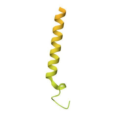
    The phosphatidylinositol 3-kinase Vps34 is part of several protein complexes. The structural organization of heterotetrameric complexes is starting to emerge, but little is known about organization of additional accessory subunits that interact with these assemblies. Combining hydrogen-deuterium exchange mass spectrometry (HDX-MS), X-ray crystallography and electron microscopy (EM), we have characterized Atg38 and its human ortholog NRBF2, accessory components of complex I consisting of Vps15-Vps34-Vps30/Atg6-Atg14 (yeast) and PIK3R4/VPS15-PIK3C3/VPS34-BECN1/Beclin 1-ATG14 (human). HDX-MS shows that Atg38 binds the Vps30-Atg14 subcomplex of complex I, using mainly its N-terminal MIT domain and bridges the coiled-coil I regions of Atg14 and Vps30 in the base of complex I. The Atg38 C-terminal domain is important for localization to the phagophore assembly site (PAS) and homodimerization. Our 2.2 Å resolution crystal structure of the Atg38 C-terminal homodimerization domain shows 2 segments of α-helices assembling into a mushroom-like asymmetric homodimer with a 4-helix cap and a parallel coiled-coil stalk. One Atg38 homodimer engages a single complex I. This is in sharp contrast to human NRBF2, which also forms a homodimer, but this homodimer can bridge 2 complex I assemblies.


  • Organizational Affiliation

    a MRC Laboratory of Molecular Biology , Cambridge , United Kingdom.


Macromolecules
Find similar proteins by:  (by identity cutoff)  |  3D Structure
Entity ID: 1
MoleculeChains Sequence LengthOrganismDetailsImage
Autophagy-related protein 38226Saccharomyces cerevisiaeMutation(s): 0 
Gene Names: ATG38YLR211CL8167.20
UniProt
Find proteins for Q05789 (Saccharomyces cerevisiae (strain ATCC 204508 / S288c))
Explore Q05789 
Go to UniProtKB:  Q05789
Entity Groups  
Sequence Clusters30% Identity50% Identity70% Identity90% Identity95% Identity100% Identity
UniProt GroupQ05789
Sequence Annotations
Expand
  • Reference Sequence
Small Molecules
Ligands 7 Unique
IDChains Name / Formula / InChI Key2D Diagram3D Interactions
IOD
Query on IOD

Download Ideal Coordinates CCD File 
UA [auth E]IODIDE ION
I
XMBWDFGMSWQBCA-UHFFFAOYSA-M
SO4
Query on SO4

Download Ideal Coordinates CCD File 
PA [auth K]SULFATE ION
O4 S
QAOWNCQODCNURD-UHFFFAOYSA-L
EDO
Query on EDO

Download Ideal Coordinates CCD File 
GB [auth I]
JB [auth J]
KB [auth J]
O [auth C]
OA [auth K]
GB [auth I],
JB [auth J],
KB [auth J],
O [auth C],
OA [auth K],
P [auth C],
QA [auth L],
Y [auth A],
Z [auth A]
1,2-ETHANEDIOL
C2 H6 O2
LYCAIKOWRPUZTN-UHFFFAOYSA-N
NO3
Query on NO3

Download Ideal Coordinates CCD File 
BA [auth B]
CA [auth G]
DA [auth G]
EA [auth G]
EB [auth I]
BA [auth B],
CA [auth G],
DA [auth G],
EA [auth G],
EB [auth I],
FA [auth G],
FB [auth I],
GA [auth H],
HB [auth J],
KA [auth K],
LA [auth K],
MA [auth K],
N [auth C],
S [auth D],
T [auth D],
W [auth A],
X [auth A],
ZA [auth F]
NITRATE ION
N O3
NHNBFGGVMKEFGY-UHFFFAOYSA-N
CL
Query on CL

Download Ideal Coordinates CCD File 
AB [auth F]
BB [auth F]
SA [auth E]
TA [auth E]
U [auth D]
AB [auth F],
BB [auth F],
SA [auth E],
TA [auth E],
U [auth D],
V [auth D]
CHLORIDE ION
Cl
VEXZGXHMUGYJMC-UHFFFAOYSA-M
NA
Query on NA

Download Ideal Coordinates CCD File 
AA [auth B]
CB [auth I]
DB [auth I]
HA [auth K]
IA [auth K]
AA [auth B],
CB [auth I],
DB [auth I],
HA [auth K],
IA [auth K],
JA [auth K],
M [auth C],
Q [auth D],
R [auth D],
RA [auth E],
VA [auth F],
WA [auth F],
XA [auth F],
YA [auth F]
SODIUM ION
Na
FKNQFGJONOIPTF-UHFFFAOYSA-N
NH4
Query on NH4

Download Ideal Coordinates CCD File 
IB [auth J],
NA [auth K]
AMMONIUM ION
H4 N
QGZKDVFQNNGYKY-UHFFFAOYSA-O
Experimental Data & Validation

Experimental Data

  • Method: X-RAY DIFFRACTION
  • Resolution: 2.20 Å
  • R-Value Free: 0.241 
  • R-Value Work: 0.205 
  • R-Value Observed: 0.206 
  • Space Group: P 21 21 2
Unit Cell:
Length ( Å )Angle ( ˚ )
a = 81.383α = 90
b = 249.385β = 90
c = 50.197γ = 90
Software Package:
Software NamePurpose
PHENIXrefinement
XDSdata reduction
Aimlessdata scaling
autoSHARPphasing

Structure Validation

View Full Validation Report


Entry History & Funding Information

Deposition Data


Funding OrganizationLocationGrant Number
Medical Research Council (United Kingdom)United KingdomMC_U105184308

Revision History  (Full details and data files)

  • Version 1.0: 2016-10-05
    Type: Initial release
  • Version 1.1: 2016-10-19
    Changes: Database references
  • Version 1.2: 2016-11-09
    Changes: Database references
  • Version 2.0: 2017-09-13
    Changes: Atomic model, Author supporting evidence, Derived calculations