Solution Binding and Structural Analyses Reveal Potential Multidrug Resistance Functions for SAV2435 and CTR107 and Other GyrI-like Proteins.
Moreno, A., Froehlig, J.R., Bachas, S., Gunio, D., Alexander, T., Vanya, A., Wade, H.(2016) Biochemistry 55: 4850-4863
- PubMed: 27505298
- DOI: https://doi.org/10.1021/acs.biochem.6b00651
- Primary Citation of Related Structures:
5KAT, 5KAU, 5KAV, 5KAW, 5KAX, 5KCB - PubMed Abstract:
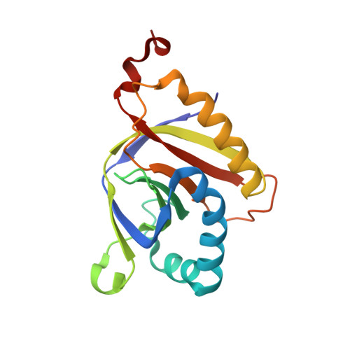
Multidrug resistance (MDR) refers to the acquired ability of cells to tolerate a broad range of toxic compounds. One mechanism cells employ is to increase the level of expression of efflux pumps for the expulsion of xenobiotics. A key feature uniting efflux-related mechanisms is multidrug (MD) recognition, either by efflux pumps themselves or by their transcriptional regulators. However, models describing MD binding by MDR effectors are incomplete, underscoring the importance of studies focused on the recognition elements and key motifs that dictate polyspecific binding. One such motif is the GyrI-like domain, which is found in several MDR proteins and is postulated to have been adapted for small-molecule binding and signaling. Here we report the solution binding properties and crystal structures of two proteins containing GyrI-like domains, SAV2435 and CTR107, bound to various ligands. Furthermore, we provide a comparison with deposited crystal structures of GyrI-like proteins, revealing key features of GyrI-like domains that not only support polyspecific binding but also are conserved among GyrI-like domains. Together, our studies suggest that GyrI-like domains perform evolutionarily conserved functions connected to multidrug binding and highlight the utility of these types of studies for elucidating mechanisms of MDR.
Organizational Affiliation:
Department of Biophysics and Biophysical Chemistry, Johns Hopkins University School of Medicine , 725 North Wolfe Street, Baltimore, Maryland 21205, United States.