5K5S

Crystal structure of the active form of human calcium-sensing receptor extracellular domain


Experimental Data Snapshot

  • Method: X-RAY DIFFRACTION
  • Resolution: 2.60 Å
  • R-Value Free: 0.222 
  • R-Value Work: 0.211 
  • R-Value Observed: 0.212 

wwPDB Validation   3D Report Full Report


Ligand Structure Quality Assessment 


This is version 1.4 of the entry. See complete history


Literature

Structural mechanism of ligand activation in human calcium-sensing receptor.

Geng, Y.Mosyak, L.Kurinov, I.Zuo, H.Sturchler, E.Cheng, T.C.Subramanyam, P.Brown, A.P.Brennan, S.C.Mun, H.C.Bush, M.Chen, Y.Nguyen, T.X.Cao, B.Chang, D.D.Quick, M.Conigrave, A.D.Colecraft, H.M.McDonald, P.Fan, Q.R.

(2016) Elife 5

  • DOI: https://doi.org/10.7554/eLife.13662
  • Primary Citation of Related Structures:  
    5K5S, 5K5T

  • PubMed Abstract: 

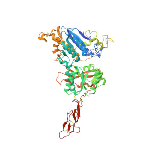
    Human calcium-sensing receptor (CaSR) is a G-protein-coupled receptor (GPCR) that maintains extracellular Ca(2+) homeostasis through the regulation of parathyroid hormone secretion. It functions as a disulfide-tethered homodimer composed of three main domains, the Venus Flytrap module, cysteine-rich domain, and seven-helix transmembrane region. Here, we present the crystal structures of the entire extracellular domain of CaSR in the resting and active conformations. We provide direct evidence that L-amino acids are agonists of the receptor. In the active structure, L-Trp occupies the orthosteric agonist-binding site at the interdomain cleft and is primarily responsible for inducing extracellular domain closure to initiate receptor activation. Our structures reveal multiple binding sites for Ca(2+) and PO4(3-) ions. Both ions are crucial for structural integrity of the receptor. While Ca(2+) ions stabilize the active state, PO4(3-) ions reinforce the inactive conformation. The activation mechanism of CaSR involves the formation of a novel dimer interface between subunits.


  • Organizational Affiliation

    Department of Pharmacology, Columbia University, New York, United States.


Macromolecules
Find similar proteins by:  (by identity cutoff)  |  3D Structure
Entity ID: 1
MoleculeChains Sequence LengthOrganismDetailsImage
Extracellular calcium-sensing receptor
A, B
615Homo sapiensMutation(s): 3 
Gene Names: CASRGPRC2APCAR1
UniProt & NIH Common Fund Data Resources
Find proteins for P41180 (Homo sapiens)
Explore P41180 
Go to UniProtKB:  P41180
PHAROS:  P41180
GTEx:  ENSG00000036828 
Entity Groups  
Sequence Clusters30% Identity50% Identity70% Identity90% Identity95% Identity100% Identity
UniProt GroupP41180
Sequence Annotations
Expand
  • Reference Sequence
Small Molecules
Ligands 4 Unique
IDChains Name / Formula / InChI Key2D Diagram3D Interactions
NAG
Query on NAG

Download Ideal Coordinates CCD File 
J [auth A],
K [auth A],
L [auth A],
M [auth A],
U [auth B]
2-acetamido-2-deoxy-beta-D-glucopyranose
C8 H15 N O6
OVRNDRQMDRJTHS-FMDGEEDCSA-N
TRP
Query on TRP

Download Ideal Coordinates CCD File 
C [auth A],
O [auth B]
TRYPTOPHAN
C11 H12 N2 O2
QIVBCDIJIAJPQS-VIFPVBQESA-N
PO4
Query on PO4

Download Ideal Coordinates CCD File 
D [auth A],
E [auth A],
P [auth B],
Q [auth B]
PHOSPHATE ION
O4 P
NBIIXXVUZAFLBC-UHFFFAOYSA-K
CA
Query on CA

Download Ideal Coordinates CCD File 
F [auth A]
G [auth A]
H [auth A]
I [auth A]
N [auth A]
F [auth A],
G [auth A],
H [auth A],
I [auth A],
N [auth A],
R [auth B],
S [auth B],
T [auth B]
CALCIUM ION
Ca
BHPQYMZQTOCNFJ-UHFFFAOYSA-N
Experimental Data & Validation

Experimental Data

  • Method: X-RAY DIFFRACTION
  • Resolution: 2.60 Å
  • R-Value Free: 0.222 
  • R-Value Work: 0.211 
  • R-Value Observed: 0.212 
  • Space Group: C 1 2 1
Unit Cell:
Length ( Å )Angle ( ˚ )
a = 107.66α = 90
b = 127.45β = 108.72
c = 146.77γ = 90
Software Package:
Software NamePurpose
BUSTERrefinement
XDSdata reduction
Aimlessdata scaling
PHASERphasing

Structure Validation

View Full Validation Report


Ligand Structure Quality Assessment 


Entry History & Funding Information

Deposition Data


Funding OrganizationLocationGrant Number
American Heart AssociationUnited States15GRNT25420002
National Institutes of Health/National Institute of General Medical Sciences (NIH/NIGMS)United StatesR01GM112973

Revision History  (Full details and data files)

  • Version 1.0: 2016-08-03
    Type: Initial release
  • Version 1.1: 2016-08-24
    Changes: Data collection
  • Version 1.2: 2017-09-20
    Changes: Author supporting evidence, Derived calculations
  • Version 1.3: 2019-12-25
    Changes: Author supporting evidence
  • Version 1.4: 2020-07-29
    Type: Remediation
    Reason: Carbohydrate remediation
    Changes: Data collection, Derived calculations, Structure summary