5K4L

Crystal structure of KDM5A in complex with a naphthyridone inhibitor


Experimental Data Snapshot

  • Method: X-RAY DIFFRACTION
  • Resolution: 3.18 Å
  • R-Value Free: 0.257 
  • R-Value Work: 0.222 
  • R-Value Observed: 0.224 

wwPDB Validation   3D Report Full Report


Ligand Structure Quality Assessment 


This is version 1.2 of the entry. See complete history


Literature

Design and evaluation of 1,7-naphthyridones as novel KDM5 inhibitors.

Labadie, S.S.Dragovich, P.S.Cummings, R.T.Deshmukh, G.Gustafson, A.Han, N.Harmange, J.C.Kiefer, J.R.Li, Y.Liang, J.Liederer, B.M.Liu, Y.Manieri, W.Mao, W.Murray, L.Ortwine, D.F.Trojer, P.VanderPorten, E.Vinogradova, M.Wen, L.

(2016) Bioorg Med Chem Lett 26: 4492-4496

  • DOI: https://doi.org/10.1016/j.bmcl.2016.07.070
  • Primary Citation of Related Structures:  
    5K4L

  • PubMed Abstract: 

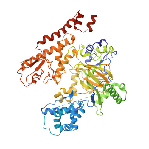
    Features from a high throughput screening (HTS) hit and a previously reported scaffold were combined to generate 1,7-naphthyridones as novel KDM5 enzyme inhibitors with nanomolar potencies. These molecules exhibited high selectivity over the related KDM4C and KDM2B isoforms. An X-ray co-crystal structure of a representative molecule bound to KDM5A showed that these inhibitors are competitive with the co-substrate (2-oxoglutarate or 2-OG).


  • Organizational Affiliation

    Genentech Inc., 1 DNA Way, South San Francisco, CA 94080, USA.


Macromolecules
Find similar proteins by:  (by identity cutoff)  |  3D Structure
Entity ID: 1
MoleculeChains Sequence LengthOrganismDetailsImage
Lysine-specific demethylase 5A
A, B
790Homo sapiensMutation(s): 0 
Gene Names: KDM5AJARID1ARBBP2RBP2
EC: 1.14.11
UniProt & NIH Common Fund Data Resources
Find proteins for P29375 (Homo sapiens)
Explore P29375 
Go to UniProtKB:  P29375
PHAROS:  P29375
GTEx:  ENSG00000073614 
Entity Groups  
Sequence Clusters30% Identity50% Identity70% Identity90% Identity95% Identity100% Identity
UniProt GroupP29375
Sequence Annotations
Expand
  • Reference Sequence

Find similar proteins by:  Sequence   |   3D Structure  

Entity ID: 2
MoleculeChains Sequence LengthOrganismDetailsImage
Unknown PeptideC [auth F],
D [auth G]
10Homo sapiensMutation(s): 0 
Gene Names: KDM5AJARID1ARBBP2RBP2
Sequence Annotations
Expand
  • Reference Sequence
Small Molecules
Ligands 3 Unique
IDChains Name / Formula / InChI Key2D Diagram3D Interactions
6QN
Query on 6QN

Download Ideal Coordinates CCD File 
H [auth A],
L [auth B]
~{N}-ethyl-4-oxidanyl-2-oxidanylidene-1~{H}-1,7-naphthyridine-3-carboxamide
C11 H11 N3 O3
RTMUDFJHLBNEDM-UHFFFAOYSA-N
ZN
Query on ZN

Download Ideal Coordinates CCD File 
F [auth A],
G [auth A],
J [auth B],
K [auth B]
ZINC ION
Zn
PTFCDOFLOPIGGS-UHFFFAOYSA-N
NI
Query on NI

Download Ideal Coordinates CCD File 
E [auth A],
I [auth B]
NICKEL (II) ION
Ni
VEQPNABPJHWNSG-UHFFFAOYSA-N
Experimental Data & Validation

Experimental Data

  • Method: X-RAY DIFFRACTION
  • Resolution: 3.18 Å
  • R-Value Free: 0.257 
  • R-Value Work: 0.222 
  • R-Value Observed: 0.224 
  • Space Group: P 31
Unit Cell:
Length ( Å )Angle ( ˚ )
a = 159.141α = 90
b = 159.141β = 90
c = 92.109γ = 120
Software Package:
Software NamePurpose
Blu-Icedata collection
PHENIXrefinement
PDB_EXTRACTdata extraction
HKL-2000data reduction
HKL-2000data scaling
PHASERphasing

Structure Validation

View Full Validation Report


Ligand Structure Quality Assessment 


Entry History 

Deposition Data

Revision History  (Full details and data files)

  • Version 1.0: 2016-08-10
    Type: Initial release
  • Version 1.1: 2016-08-24
    Changes: Database references
  • Version 1.2: 2016-09-07
    Changes: Database references