


Crystal structure of Retinoic acid receptor-related orphan receptor (ROR) gamma ligand binding domain
Lu, J., Rastinejad, F.To be published.
Experimental Data Snapshot
wwPDB Validation 3D Report Full Report

Entity ID: 1 | |||||
|---|---|---|---|---|---|
| Molecule | Chains | Sequence Length | Organism | Details | Image |
| Nuclear receptor ROR-gamma | 247 | Homo sapiens | Mutation(s): 0 Gene Names: RORC, NR1F3, RORG, RZRG | 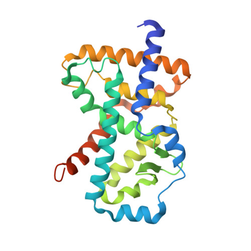 | |
UniProt & NIH Common Fund Data Resources | |||||
Find proteins for P51449 (Homo sapiens) Explore P51449 Go to UniProtKB: P51449 | |||||
PHAROS: P51449 GTEx: ENSG00000143365 | |||||
Entity Groups | |||||
| Sequence Clusters | 30% Identity50% Identity70% Identity90% Identity95% Identity100% Identity | ||||
| UniProt Group | P51449 | ||||
Sequence AnnotationsExpand | |||||
| |||||
| Length ( Å ) | Angle ( ˚ ) |
|---|---|
| a = 99.346 | α = 90 |
| b = 99.346 | β = 90 |
| c = 129.411 | γ = 120 |
| Software Name | Purpose |
|---|---|
| REFMAC | refinement |
| HKL-2000 | data collection |
| PDB_EXTRACT | data extraction |
| HKL-3000 | data reduction |
| HKL-3000 | data scaling |
| PHASER | phasing |
RCSB PDB (citation) is hosted by
RCSB PDB is a member of the

