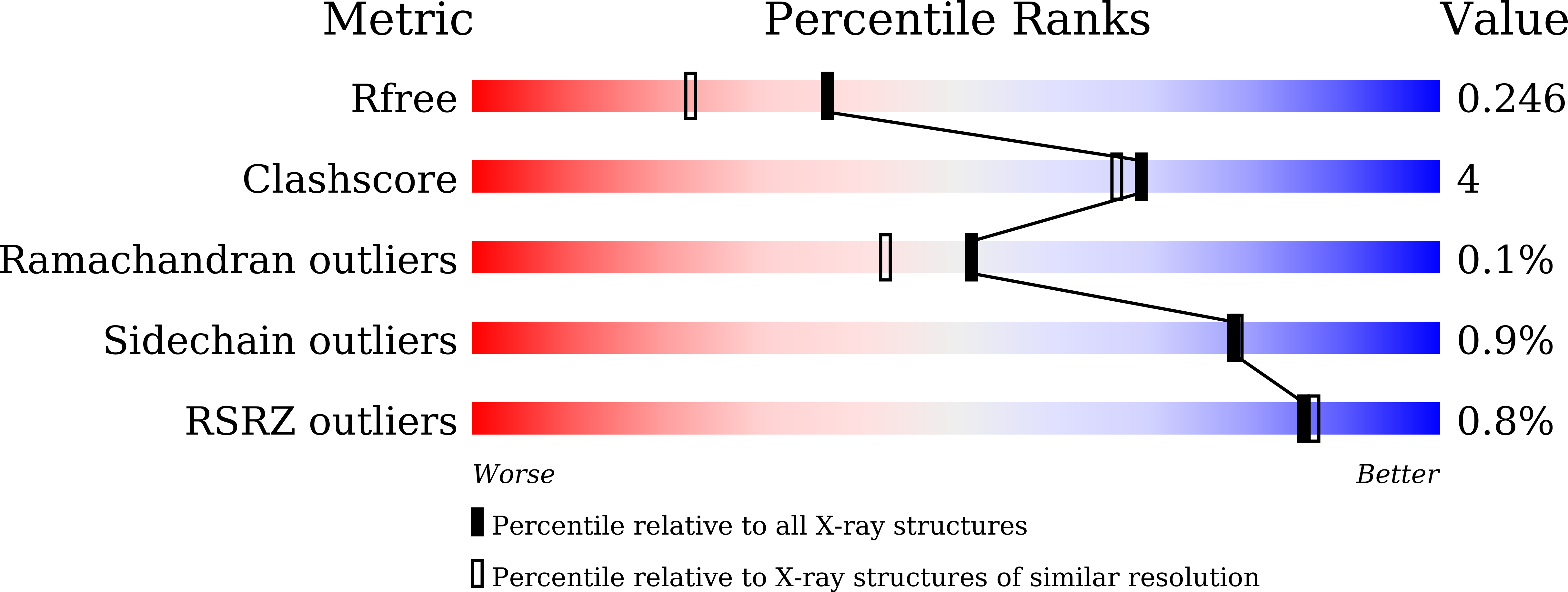
Crystal structure of peroxiredoxin 3 fromVibrio vulnificusand its implications for scavenging peroxides and nitric oxide.
Ahn, J., Jang, K.K., Jo, I., Nurhasni, H., Lim, J.G., Yoo, J.W., Choi, S.H., Ha, N.C.(2018) IUCrJ 5: 82-92
- PubMed: 29354274
- DOI: https://doi.org/10.1107/S205225251701750X
- Primary Citation of Related Structures:
5K1G, 5K2I, 5K2J - PubMed Abstract:
Peroxiredoxins (Prxs) are ubiquitous cysteine-based peroxidase enzymes. Recently, a new type of Prx, Vv Prx3, was identified in the pathogenic bacterium Vibrio vulnificus as being important for survival in macrophages. It employs only one catalytic cysteine residue to decompose peroxides. Here, crystal structures of Vv Prx3 representing its reduced and oxidized states have been determined, together with an H 2 O 2 -bound structure, at high resolution. The crystal structure representing the reduced Prx3 showed a typical dimeric interface, called the A-type interface. However, Vv Prx3 forms an oligomeric interface mediated by a disulfide bond between two catalytic cysteine residues from two adjacent dimers, which differs from the doughnut-like oligomers that appear in most Prxs. Subsequent biochemical studies showed that this disulfide bond was induced by treatment with nitric oxide (NO) as well as with peroxides. Consistently, NO treatment induced expression of the prx3 gene in V. vulnificus , and Vv Prx3 was crucial for the survival of bacteria in the presence of NO. Taken together, the function and mechanism of Vv Prx3 in scavenging peroxides and NO stress via oligomerization are proposed. These findings contribute to the understanding of the diverse functions of Prxs during pathogenic processes at the molecular level.
Organizational Affiliation:
Department of Agricultural Biotechnology, Seoul National University, 1 Gwanak-ro, Seoul 08826, Republic of Korea.


