5K16

Crystal structure of free Ubiquitin-specific protease 12


Experimental Data Snapshot

  • Method: X-RAY DIFFRACTION
  • Resolution: 2.60 Å
  • R-Value Free: 0.246 
  • R-Value Work: 0.190 
  • R-Value Observed: 0.193 

wwPDB Validation   3D Report Full Report


This is version 1.4 of the entry. See complete history


Literature

Allosteric Activation of Ubiquitin-Specific Proteases by beta-Propeller Proteins UAF1 and WDR20.

Li, H.Lim, K.S.Kim, H.Hinds, T.R.Jo, U.Mao, H.Weller, C.E.Sun, J.Chatterjee, C.D'Andrea, A.D.Zheng, N.

(2016) Mol Cell 63: 249-260

  • DOI: https://doi.org/10.1016/j.molcel.2016.05.031
  • Primary Citation of Related Structures:  
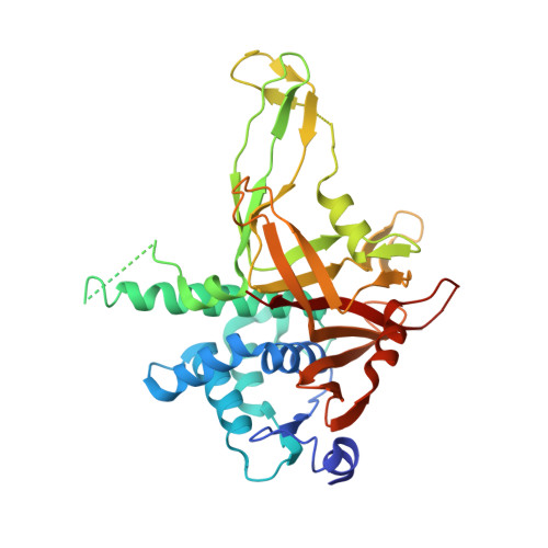
    5K16, 5K19, 5K1A, 5K1B, 5K1C

  • PubMed Abstract: 

    Ubiquitin-specific proteases (USPs) constitute the largest family of deubiquitinating enzymes, whose catalytic competency is often modulated by their binding partners through unknown mechanisms. Here we report on a series of crystallographic and biochemical analyses of an evolutionarily conserved deubiquitinase, USP12, which is activated by two β-propeller proteins, UAF1 and WDR20. Our structures reveal that UAF1 and WDR20 interact with USP12 at two distinct sites far from its catalytic center. Without increasing the substrate affinity of USP12, the two β-propeller proteins potentiate the enzyme through different allosteric mechanisms. UAF1 docks at the distal end of the USP12 Fingers domain and induces a cascade of structural changes that reach a critical ubiquitin-contacting loop adjacent to the catalytic cleft. By contrast, WDR20 anchors at the base of this loop and remotely modulates the catalytic center of the enzyme. Our results provide a mechanistic example for allosteric activation of USPs by their regulatory partners.


  • Organizational Affiliation

    Department of Pharmacology, University of Washington, Seattle, WA 98195, USA; Howard Hughes Medical Institute, Box 357280, University of Washington, Seattle, WA 98195, USA.


Macromolecules
Find similar proteins by:  (by identity cutoff)  |  3D Structure
Entity ID: 1
MoleculeChains Sequence LengthOrganismDetailsImage
Ubiquitin carboxyl-terminal hydrolase 12
A, B
355Homo sapiensMutation(s): 0 
Gene Names: USP12UBH1USP12L1
EC: 3.4.19.12
UniProt & NIH Common Fund Data Resources
Find proteins for O75317 (Homo sapiens)
Explore O75317 
Go to UniProtKB:  O75317
PHAROS:  O75317
GTEx:  ENSG00000152484 
Entity Groups  
Sequence Clusters30% Identity50% Identity70% Identity90% Identity95% Identity100% Identity
UniProt GroupO75317
Sequence Annotations
Expand
  • Reference Sequence
Experimental Data & Validation

Experimental Data

  • Method: X-RAY DIFFRACTION
  • Resolution: 2.60 Å
  • R-Value Free: 0.246 
  • R-Value Work: 0.190 
  • R-Value Observed: 0.193 
  • Space Group: P 21 21 21
Unit Cell:
Length ( Å )Angle ( ˚ )
a = 52.396α = 90
b = 109.636β = 90
c = 134.193γ = 90
Software Package:
Software NamePurpose
PHENIXrefinement
PHENIXphasing
Cootmodel building
HKL-2000data reduction
HKL-2000data scaling

Structure Validation

View Full Validation Report


Entry History & Funding Information

Deposition Data


Funding OrganizationLocationGrant Number
Howard Hughes Medical Institute (HHMI)United States--

Revision History  (Full details and data files)

  • Version 1.0: 2016-07-20
    Type: Initial release
  • Version 1.1: 2016-08-10
    Changes: Database references
  • Version 1.2: 2017-11-01
    Changes: Author supporting evidence, Database references, Derived calculations
  • Version 1.3: 2019-11-20
    Changes: Author supporting evidence
  • Version 1.4: 2023-09-27
    Changes: Data collection, Database references, Refinement description