5K13

Crystal structure of the RAR alpha ligand-binding domain in complex with an antagonist


Experimental Data Snapshot

  • Method: X-RAY DIFFRACTION
  • Resolution: 1.85 Å
  • R-Value Free: 0.222 
  • R-Value Work: 0.195 
  • R-Value Observed: 0.196 

wwPDB Validation   3D Report Full Report


Ligand Structure Quality Assessment 


This is version 1.1 of the entry. See complete history


Literature

Identification of potent and selective retinoic acid receptor gamma (RAR gamma ) antagonists for the treatment of osteoarthritis pain using structure based drug design.

Hughes, N.E.Bleisch, T.J.Jones, S.A.Richardson, T.I.Doti, R.A.Wang, Y.Stout, S.L.Durst, G.L.Chambers, M.G.Oskins, J.L.Lin, C.Adams, L.A.Page, T.J.Barr, R.J.Zink, R.W.Osborne, H.Montrose-Rafizadeh, C.Norman, B.H.

(2016) Bioorg Med Chem Lett 26: 3274-3277

  • DOI: https://doi.org/10.1016/j.bmcl.2016.05.056
  • Primary Citation of Related Structures:  
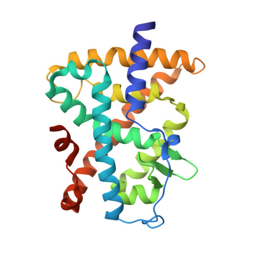
    5K13

  • PubMed Abstract: 

    A series of triaryl pyrazoles were identified as potent pan antagonists for the retinoic acid receptors (RARs) α, β and γ. X-ray crystallography and structure-based drug design were used to improve selectivity for RARγ by targeting residue differences in the ligand binding pockets of these receptors. This resulted in the discovery of novel antagonists which maintained RARγ potency but were greater than 500-fold selective versus RARα and RARβ. The potent and selective RARγ antagonist LY2955303 demonstrated good pharmacokinetic properties and was efficacious in the MIA model of osteoarthritis-like joint pain. This compound demonstrated an improved margin to RARα-mediated adverse effects.


  • Organizational Affiliation

    Discovery Chemistry Research and Technologies, Eli Lilly and Company, Lilly Corporate Center, Indianapolis, IN 46285, United States.


Macromolecules
Find similar proteins by:  (by identity cutoff)  |  3D Structure
Entity ID: 1
MoleculeChains Sequence LengthOrganismDetailsImage
Retinoic acid receptor alpha246Homo sapiensMutation(s): 0 
Gene Names: RARANR1B1
UniProt & NIH Common Fund Data Resources
Find proteins for P10276 (Homo sapiens)
Explore P10276 
Go to UniProtKB:  P10276
PHAROS:  P10276
GTEx:  ENSG00000131759 
Entity Groups  
Sequence Clusters30% Identity50% Identity70% Identity90% Identity95% Identity100% Identity
UniProt GroupP10276
Sequence Annotations
Expand
  • Reference Sequence
Small Molecules
Ligands 1 Unique
IDChains Name / Formula / InChI Key2D Diagram3D Interactions
6Q7
Query on 6Q7

Download Ideal Coordinates CCD File 
B [auth A]4-{5-(3-tert-butylphenyl)-1-[4-(methylsulfonyl)phenyl]-1H-pyrazol-3-yl}benzoic acid
C27 H26 N2 O4 S
HPXXLUZXJROKDE-UHFFFAOYSA-N
Binding Affinity Annotations 
IDSourceBinding Affinity
6Q7 Binding MOAD:  5K13 Ki: 1.8 (nM) from 1 assay(s)
BindingDB:  5K13 Ki: 1.8 (nM) from 1 assay(s)
Experimental Data & Validation

Experimental Data

  • Method: X-RAY DIFFRACTION
  • Resolution: 1.85 Å
  • R-Value Free: 0.222 
  • R-Value Work: 0.195 
  • R-Value Observed: 0.196 
  • Space Group: C 1 2 1
Unit Cell:
Length ( Å )Angle ( ˚ )
a = 88.496α = 90
b = 62.541β = 106.05
c = 49.347γ = 90
Software Package:
Software NamePurpose
SCALAdata scaling
BUSTER-TNTrefinement
PDB_EXTRACTdata extraction
MOSFLMdata reduction
MOLREPphasing

Structure Validation

View Full Validation Report


Ligand Structure Quality Assessment 


Entry History 

Deposition Data

Revision History  (Full details and data files)

  • Version 1.0: 2016-06-22
    Type: Initial release
  • Version 1.1: 2023-09-27
    Changes: Data collection, Database references, Derived calculations, Refinement description