5JZX

Crystal Structure of UDP-N-acetylenolpyruvoylglucosamine reductase (MurB) from Mycobacterium tuberculosis


Experimental Data Snapshot

  • Method: X-RAY DIFFRACTION
  • Resolution: 2.20 Å
  • R-Value Free: 0.259 
  • R-Value Work: 0.207 
  • R-Value Observed: 0.210 

wwPDB Validation   3D Report Full Report


Ligand Structure Quality Assessment 


This is version 1.3 of the entry. See complete history


Literature

Crystal structure of UDP-N-acetylglucosamine-enolpyruvate reductase (MurB) from Mycobacterium tuberculosis

Eniyan, K.Dharavath, S.Vijayan, R.Bajpai, U.Gourinath, S.

(2017) Biochim Biophys Acta 1866: 397-406

  • DOI: https://doi.org/10.1016/j.bbapap.2017.11.013
  • Primary Citation of Related Structures:  
    5JZX

  • PubMed Abstract: 

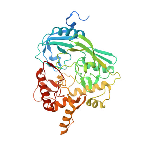
    The biosynthesis of UDP-N-acetylmuramic acid (UDP-MurNAc) by reduction of UDP-N-acetylglucosamine-enolpyruvate (UDP-GlcNAc-EP) in an NADPH and FAD-dependent reaction in bacteria is one of the key steps in peptidoglycan biosynthesis catalyzed by UDP-N-acetylglucosamine-enolpyruvate reductase (MurB). Here, we present the crystal structure of Mycobacterium tuberculosis MurB (MtbMurB) with FAD as the prosthetic group at 2.0Å resolution. There are six molecules in asymmetric unit in the form of dimers. Each protomer can be subdivided into three domains and the prosthetic group, FAD is bound in the active site between domain I and domain II. Comparison of MtbMurB structure with the structures of the Escherichia coli MurB (in complex with UDP-GlcNAc-EP) and Pseudomonas aeruginosa MurB (in complex with NADPH) showed all three structures share similar domain architecture and residues in the active site. The nicotinamide and the enol pyruvyl moieties are well aligned upon superimposition, both positioned in suitable position for hydride transfer to and from FAD. The comparison studies and MD simulations demonstrate that the two lobes of domain-III become more flexible. The substrates (NADPH and UDP-GlcNAc-EP) binding responsible for open conformation of MurB, suggesting that NADPH and UDP-GlcNAc-EP interactions are conformationally stable. Our findings provide a detail mechanism about the closed to open state by binding of NADPH and UDP-GlcNAc-EP induces the conformational changes of MurB structure that may trigger the MurB catalytic reaction.


  • Organizational Affiliation

    Department of Biomedical Science, Acharya Narendra Dev College, University of Delhi, New Delhi, India.


Macromolecules
Find similar proteins by:  (by identity cutoff)  |  3D Structure
Entity ID: 1
MoleculeChains Sequence LengthOrganismDetailsImage
UDP-N-acetylenolpyruvoylglucosamine reductase
A, B, C, D, E
A, B, C, D, E, F
405Mycobacterium tuberculosis H37RvMutation(s): 0 
Gene Names: murB
EC: 1.3.1.98
UniProt
Find proteins for P9WJL9 (Mycobacterium tuberculosis (strain ATCC 25618 / H37Rv))
Explore P9WJL9 
Go to UniProtKB:  P9WJL9
Entity Groups  
Sequence Clusters30% Identity50% Identity70% Identity90% Identity95% Identity100% Identity
UniProt GroupP9WJL9
Sequence Annotations
Expand
  • Reference Sequence
Small Molecules
Ligands 2 Unique
IDChains Name / Formula / InChI Key2D Diagram3D Interactions
FAD
Query on FAD

Download Ideal Coordinates CCD File 
G [auth A]
I [auth B]
K [auth C]
M [auth D]
N [auth E]
G [auth A],
I [auth B],
K [auth C],
M [auth D],
N [auth E],
O [auth F]
FLAVIN-ADENINE DINUCLEOTIDE
C27 H33 N9 O15 P2
VWWQXMAJTJZDQX-UYBVJOGSSA-N
K
Query on K

Download Ideal Coordinates CCD File 
H [auth A],
J [auth B],
L [auth C]
POTASSIUM ION
K
NPYPAHLBTDXSSS-UHFFFAOYSA-N
Experimental Data & Validation

Experimental Data

  • Method: X-RAY DIFFRACTION
  • Resolution: 2.20 Å
  • R-Value Free: 0.259 
  • R-Value Work: 0.207 
  • R-Value Observed: 0.210 
  • Space Group: C 1 2 1
Unit Cell:
Length ( Å )Angle ( ˚ )
a = 274.924α = 90
b = 79.966β = 112.44
c = 138.996γ = 90
Software Package:
Software NamePurpose
REFMACrefinement
HKL-2000data reduction
HKL-2000data scaling
PHASERphasing

Structure Validation

View Full Validation Report


Ligand Structure Quality Assessment 


Entry History 

Deposition Data

Revision History  (Full details and data files)

  • Version 1.0: 2017-05-10
    Type: Initial release
  • Version 1.1: 2017-12-27
    Changes: Database references
  • Version 1.2: 2018-01-17
    Changes: Database references
  • Version 1.3: 2023-11-08
    Changes: Data collection, Database references, Derived calculations, Refinement description