5JZN

Crystal structure of DCLK1-KD in complex with NVP-TAE684


Experimental Data Snapshot

  • Method: X-RAY DIFFRACTION
  • Resolution: 2.85 Å
  • R-Value Free: 0.225 
  • R-Value Work: 0.181 
  • R-Value Observed: 0.183 

wwPDB Validation   3D Report Full Report


Ligand Structure Quality Assessment 


This is version 1.2 of the entry. See complete history


Literature

Biochemical and Structural Insights into Doublecortin-like Kinase Domain 1.

Patel, O.Dai, W.Mentzel, M.Griffin, M.D.Serindoux, J.Gay, Y.Fischer, S.Sterle, S.Kropp, A.Burns, C.J.Ernst, M.Buchert, M.Lucet, I.S.

(2016) Structure 24: 1550-1561

  • DOI: https://doi.org/10.1016/j.str.2016.07.008
  • Primary Citation of Related Structures:  
    5JZJ, 5JZN

  • PubMed Abstract: 

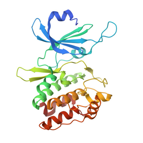
    Doublecortin-like kinase 1 (DCLK1) is a serine/threonine kinase that belongs to the family of microtubule-associated proteins. Originally identified for its role in neurogenesis, DCLK1 has recently been shown to regulate biological processes outside of the CNS. DCLK1 is among the 15 most common putative driver genes for gastric cancers and is highly mutated across various other human cancers. However, our present understanding of how DCLK1 dysfunction leads to tumorigenesis is limited. Here, we provide evidence that DCLK1 kinase activity negatively regulates microtubule polymerization. We present the crystal structure of the DCLK1 kinase domain at 1.7 Å resolution, providing detailed insight into the ATP-binding site that will serve as a framework for future drug design. This structure also allowed for the mapping of cancer-causing mutations within the kinase domain, suggesting that a loss of kinase function may contribute to tumorigenesis.


  • Organizational Affiliation

    The Walter and Eliza Hall Institute of Medical Research, Parkville, VIC 3052, Australia; Department of Medical Biology, University of Melbourne, Parkville, VIC 3052, Australia. Electronic address: patel.o@wehi.edu.au.


Macromolecules
Find similar proteins by:  (by identity cutoff)  |  3D Structure
Entity ID: 1
MoleculeChains Sequence LengthOrganismDetailsImage
Serine/threonine-protein kinase DCLK1
A, B
280Homo sapiensMutation(s): 0 
Gene Names: DCLK1DCAMKL1DCDC3AKIAA0369
EC: 2.7.11.1
UniProt & NIH Common Fund Data Resources
Find proteins for O15075 (Homo sapiens)
Explore O15075 
Go to UniProtKB:  O15075
PHAROS:  O15075
GTEx:  ENSG00000133083 
Entity Groups  
Sequence Clusters30% Identity50% Identity70% Identity90% Identity95% Identity100% Identity
UniProt GroupO15075
Sequence Annotations
Expand
  • Reference Sequence
Small Molecules
Ligands 2 Unique
IDChains Name / Formula / InChI Key2D Diagram3D Interactions
GUI
Query on GUI

Download Ideal Coordinates CCD File 
C [auth A],
E [auth B]
5-CHLORO-N-[2-METHOXY-4-[4-(4-METHYLPIPERAZIN-1-YL)PIPERIDIN-1-YL]PHENYL]-N'-(2-PROPAN-2-YLSULFONYLPHENYL)PYRIMIDINE-2,4-DIAMINE
C30 H40 Cl N7 O3 S
QQWUGDVOUVUTOY-UHFFFAOYSA-N
SO4
Query on SO4

Download Ideal Coordinates CCD File 
D [auth A]SULFATE ION
O4 S
QAOWNCQODCNURD-UHFFFAOYSA-L
Experimental Data & Validation

Experimental Data

  • Method: X-RAY DIFFRACTION
  • Resolution: 2.85 Å
  • R-Value Free: 0.225 
  • R-Value Work: 0.181 
  • R-Value Observed: 0.183 
  • Space Group: P 61
Unit Cell:
Length ( Å )Angle ( ˚ )
a = 106.36α = 90
b = 106.36β = 90
c = 157.37γ = 120
Software Package:
Software NamePurpose
Aimlessdata scaling
PHENIXrefinement
PDB_EXTRACTdata extraction
MOSFLMdata reduction
PHASERphasing

Structure Validation

View Full Validation Report


Ligand Structure Quality Assessment 


Entry History 

Deposition Data

Revision History  (Full details and data files)

  • Version 1.0: 2016-08-24
    Type: Initial release
  • Version 1.1: 2017-08-30
    Changes: Data collection, Database references, Derived calculations
  • Version 1.2: 2023-09-27
    Changes: Data collection, Database references, Refinement description