5JZ9

Crystal structure of HsaD bound to 3,5-dichloro-4-hydroxybenzenesulphonic acid


Experimental Data Snapshot

  • Method: X-RAY DIFFRACTION
  • Resolution: 2.68 Å
  • R-Value Free: 0.240 
  • R-Value Work: 0.213 
  • R-Value Observed: 0.214 

wwPDB Validation   3D Report Full Report


Ligand Structure Quality Assessment 


This is version 1.3 of the entry. See complete history


Literature

Investigation of the mycobacterial enzyme HsaD as a potential novel target for anti-tubercular agents using a fragment-based drug design approach.

Ryan, A.Polycarpou, E.Lack, N.A.Evangelopoulos, D.Sieg, C.Halman, A.Bhakta, S.Eleftheriadou, O.McHugh, T.D.Keany, S.Lowe, E.D.Ballet, R.Abuhammad, A.Jacobs, W.R.Ciulli, A.Sim, E.

(2017) Br J Pharmacol 174: 2209-2224

  • DOI: https://doi.org/10.1111/bph.13810
  • Primary Citation of Related Structures:  
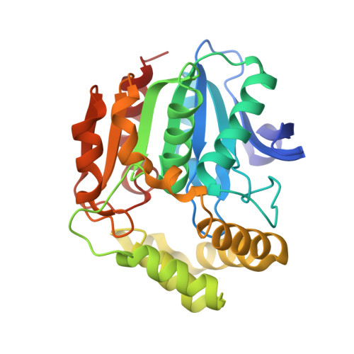
    5JZ9, 5JZB, 5JZS

  • PubMed Abstract: 

    With the emergence of extensively drug-resistant tuberculosis, there is a need for new anti-tubercular drugs that work through novel mechanisms of action. The meta cleavage product hydrolase, HsaD, has been demonstrated to be critical for the survival of Mycobacterium tuberculosis in macrophages and is encoded in an operon involved in cholesterol catabolism, which is identical in M. tuberculosis and M. bovis BCG.


  • Organizational Affiliation

    Faculty of Science, Engineering and Computing, Kingston University London, Kingston upon Thames, UK.


Macromolecules
Find similar proteins by:  (by identity cutoff)  |  3D Structure
Entity ID: 1
MoleculeChains Sequence LengthOrganismDetailsImage
4,5:9,10-diseco-3-hydroxy-5,9,17-trioxoandrosta-1(10),2-diene-4-oate hydrolase
A, B
284Mycobacterium tuberculosis CDC1551Mutation(s): 0 
Gene Names: hsaDbphDMT3674
EC: 3.7.1.17 (PDB Primary Data), 3.7.1.8 (PDB Primary Data)
UniProt
Find proteins for P9WNH5 (Mycobacterium tuberculosis (strain ATCC 25618 / H37Rv))
Explore P9WNH5 
Go to UniProtKB:  P9WNH5
Entity Groups  
Sequence Clusters30% Identity50% Identity70% Identity90% Identity95% Identity100% Identity
UniProt GroupP9WNH5
Sequence Annotations
Expand
  • Reference Sequence
Small Molecules
Ligands 1 Unique
IDChains Name / Formula / InChI Key2D Diagram3D Interactions
6OR
Query on 6OR

Download Ideal Coordinates CCD File 
C [auth A],
D [auth B]
3,5-dichloro-4-hydroxybenzene-1-sulfonic acid
C6 H4 Cl2 O4 S
PGDCAFRJYQICAY-UHFFFAOYSA-N
Experimental Data & Validation

Experimental Data

  • Method: X-RAY DIFFRACTION
  • Resolution: 2.68 Å
  • R-Value Free: 0.240 
  • R-Value Work: 0.213 
  • R-Value Observed: 0.214 
  • Space Group: I 21 21 21
Unit Cell:
Length ( Å )Angle ( ˚ )
a = 81.73α = 90
b = 82.318β = 90
c = 193.584γ = 90
Software Package:
Software NamePurpose
Aimlessdata scaling
PHASERphasing
PHENIXrefinement
PDB_EXTRACTdata extraction
XDSdata reduction

Structure Validation

View Full Validation Report


Ligand Structure Quality Assessment 


Entry History 

Revision History  (Full details and data files)

  • Version 1.0: 2017-04-05
    Type: Initial release
  • Version 1.1: 2017-04-12
    Changes: Database references
  • Version 1.2: 2017-07-05
    Changes: Database references
  • Version 1.3: 2024-01-10
    Changes: Advisory, Data collection, Database references, Refinement description