5JVO

Crystal structure of the Arginine Repressor from the pathogenic bacterium Corynebacterium pseudotuberculosis


Experimental Data Snapshot

  • Method: X-RAY DIFFRACTION
  • Resolution: 1.90 Å
  • R-Value Free: 0.225 
  • R-Value Work: 0.180 
  • R-Value Observed: 0.182 

wwPDB Validation   3D Report Full Report


Ligand Structure Quality Assessment 


This is version 1.1 of the entry. See complete history


Literature

Tyrosine binding and promiscuity in the arginine repressor from the pathogenic bacterium Corynebacterium pseudotuberculosis.

Mariutti, R.B.Ullah, A.Araujo, G.C.Murakami, M.T.Arni, R.K.

(2016) Biochem Biophys Res Commun 475: 350-355

  • DOI: https://doi.org/10.1016/j.bbrc.2016.05.091
  • Primary Citation of Related Structures:  
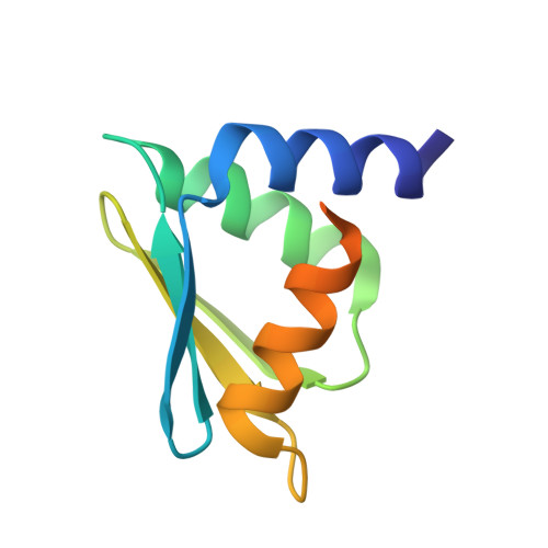
    5JVO

  • PubMed Abstract: 

    The arginine repressor (ArgR) regulates arginine biosynthesis in a number of microorganisms and consists of two domains interlinked by a short peptide; the N-terminal domain is involved in DNA binding and the C-terminal domain binds arginine and forms a hexamer made-up of a dimer of trimers. The crystal structure of the C-terminal domain of ArgR from the pathogenic Corynebacterium pseudotuberculosis determined at 1.9 Å resolution contains a tightly bound tyrosine at the arginine-binding site indicating hitherto unobserved promiscuity. Structural analysis of the binding pocket displays clear molecular adaptations to accommodate tyrosine binding suggesting the possible existence of an alternative regulatory process in this pathogenic bacterium.


  • Organizational Affiliation

    Multiuser Center for Biomolecular Innovation, IBILCE/UNESP, São José do Rio Preto, SP, 15054-000, Brazil. Electronic address: mariutti@ibilce.unesp.br.


Macromolecules
Find similar proteins by:  (by identity cutoff)  |  3D Structure
Entity ID: 1
MoleculeChains Sequence LengthOrganismDetailsImage
Arginine repressor
A, B
97Corynebacterium pseudotuberculosisMutation(s): 0 
Gene Names: argRCpE19_0967
UniProt
Find proteins for D9QA55 (Corynebacterium pseudotuberculosis (strain C231))
Explore D9QA55 
Go to UniProtKB:  D9QA55
Entity Groups  
Sequence Clusters30% Identity50% Identity70% Identity90% Identity95% Identity100% Identity
UniProt GroupD9QA55
Sequence Annotations
Expand
  • Reference Sequence
Experimental Data & Validation

Experimental Data

  • Method: X-RAY DIFFRACTION
  • Resolution: 1.90 Å
  • R-Value Free: 0.225 
  • R-Value Work: 0.180 
  • R-Value Observed: 0.182 
  • Space Group: P 21 3
Unit Cell:
Length ( Å )Angle ( ˚ )
a = 83.191α = 90
b = 83.191β = 90
c = 83.191γ = 90
Software Package:
Software NamePurpose
XSCALEdata scaling
REFMACrefinement
PDB_EXTRACTdata extraction
XDSdata reduction
PHASERphasing

Structure Validation

View Full Validation Report


Ligand Structure Quality Assessment 


Entry History 

Deposition Data

Revision History  (Full details and data files)

  • Version 1.0: 2016-08-31
    Type: Initial release
  • Version 1.1: 2023-09-27
    Changes: Data collection, Database references, Derived calculations, Refinement description