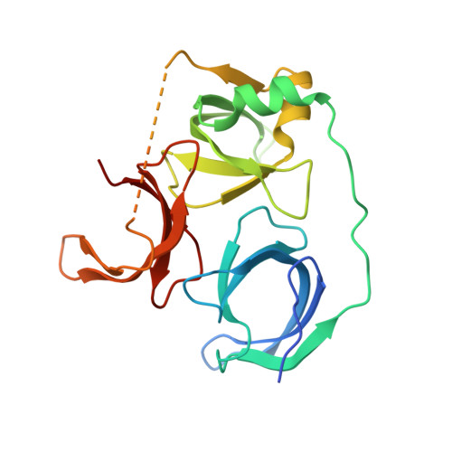
5JSJ

Crystal structure of Spindlin1 bound to compound EML631


Experimental Data Snapshot

  • Method: X-RAY DIFFRACTION
  • Resolution: 2.35 Å
  • R-Value Free: 0.249 
  • R-Value Work: 0.199 
  • R-Value Observed: 0.201 

wwPDB Validation   3D Report Full Report


Ligand Structure Quality Assessment 


This is version 1.2 of the entry. See complete history


Literature

Developing Spindlin1 small-molecule inhibitors by using protein microarrays

Bae, N.Viviano, M.Su, X.Lv, J.Cheng, D.Sagum, C.Castellano, S.Bai, X.Johnson, C.Khalil, M.I.Shen, J.Chen, K.Li, H.Sbardella, G.Bedford, M.T.

(2017) Nat Chem Biol 13: 750-756

  • DOI: https://doi.org/10.1038/nchembio.2377
  • Primary Citation of Related Structures:  
    5JSG, 5JSJ

  • PubMed Abstract: 

    The discovery of inhibitors of methyl- and acetyl-binding domains has provided evidence for the 'druggability' of epigenetic effector molecules. The small-molecule probe UNC1215 prevents methyl-dependent protein-protein interactions by engaging the aromatic cage of MBT domains and, with lower affinity, Tudor domains. Using a library of tagged UNC1215 analogs, we screened a protein-domain microarray of human methyllysine effector molecules to rapidly detect compounds with new binding profiles with either increased or decreased specificity. Using this approach, we identified a compound (EML405) that acquired a novel interaction with the Tudor-domain-containing protein Spindlin1 (SPIN1). Structural studies facilitated the rational synthesis of SPIN1 inhibitors with increased selectivity (EML631-633), which engage SPIN1 in cells, block its ability to 'read' H3K4me3 marks and inhibit its transcriptional-coactivator activity. Protein microarrays can thus be used as a platform to 'target-hop' and identify small molecules that bind and compete with domain-motif interactions.


  • Organizational Affiliation

    Department of Epigenetics and Molecular Carcinogenesis, University of Texas MD Anderson Cancer Center, Smithville, Texas, USA.


Macromolecules
Find similar proteins by:  (by identity cutoff)  |  3D Structure
Entity ID: 1
MoleculeChains Sequence LengthOrganismDetailsImage
Spindlin-1
A, B
222Homo sapiensMutation(s): 0 
Gene Names: SPIN1OCRSPIN
UniProt & NIH Common Fund Data Resources
Find proteins for Q9Y657 (Homo sapiens)
Explore Q9Y657 
Go to UniProtKB:  Q9Y657
PHAROS:  Q9Y657
GTEx:  ENSG00000106723 
Entity Groups  
Sequence Clusters30% Identity50% Identity70% Identity90% Identity95% Identity100% Identity
UniProt GroupQ9Y657
Sequence Annotations
Expand
  • Reference Sequence
Small Molecules
Binding Affinity Annotations 
IDSourceBinding Affinity
6PD Binding MOAD:  5JSJ Kd: 4000 (nM) from 1 assay(s)
Experimental Data & Validation

Experimental Data

  • Method: X-RAY DIFFRACTION
  • Resolution: 2.35 Å
  • R-Value Free: 0.249 
  • R-Value Work: 0.199 
  • R-Value Observed: 0.201 
  • Space Group: P 1 21 1
Unit Cell:
Length ( Å )Angle ( ˚ )
a = 42.79α = 90
b = 123.827β = 92.22
c = 49.663γ = 90
Software Package:
Software NamePurpose
PHENIXrefinement
HKL-2000data scaling
PDB_EXTRACTdata extraction
HKL-2000data reduction
MOLREPphasing

Structure Validation

View Full Validation Report


Ligand Structure Quality Assessment 


Entry History 

Deposition Data

  • Released Date: 2017-05-24 
  • Deposition Author(s): Su, X., Li, H.

Revision History  (Full details and data files)

  • Version 1.0: 2017-05-24
    Type: Initial release
  • Version 1.1: 2017-07-26
    Changes: Database references
  • Version 1.2: 2024-03-20
    Changes: Data collection, Database references, Derived calculations