5JQK

The Xray Crystal Structure of P. falciparum Aminopeptidase P


Experimental Data Snapshot

  • Method: X-RAY DIFFRACTION
  • Resolution: 2.35 Å
  • R-Value Free: 0.226 
  • R-Value Work: 0.189 
  • R-Value Observed: 0.190 

wwPDB Validation   3D Report Full Report


This is version 1.4 of the entry. See complete history


Literature

Structure and substrate fingerprint of aminopeptidase P from Plasmodium falciparum.

Drinkwater, N.Sivaraman, K.K.Bamert, R.S.Rut, W.Mohamed, K.Vinh, N.B.Scammells, P.J.Drag, M.McGowan, S.

(2016) Biochem J 473: 3189-3204

  • DOI: https://doi.org/10.1042/BCJ20160550
  • Primary Citation of Related Structures:  
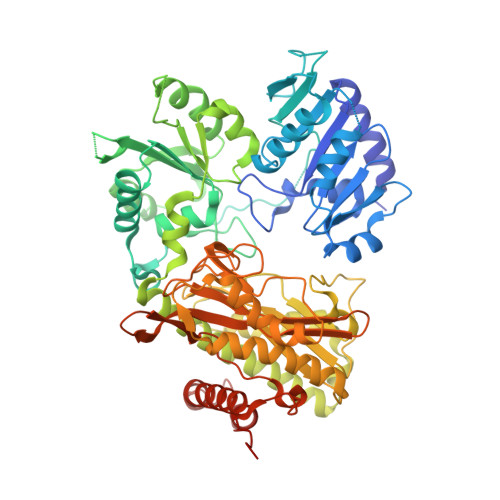
    5JQK, 5JR6

  • PubMed Abstract: 

    Malaria is one of the world's most prevalent parasitic diseases, with over 200 million cases annually. Alarmingly, the spread of drug-resistant parasites threatens the effectiveness of current antimalarials and has made the development of novel therapeutic strategies a global health priority. Malaria parasites have a complicated lifecycle, involving an asymptomatic 'liver stage' and a symptomatic 'blood stage'. During the blood stage, the parasites utilise a proteolytic cascade to digest host hemoglobin, which produces free amino acids absolutely necessary for parasite growth and reproduction. The enzymes required for hemoglobin digestion are therefore attractive therapeutic targets. The final step of the cascade is catalyzed by several metalloaminopeptidases, including aminopeptidase P (APP). We developed a novel platform to examine the substrate fingerprint of APP from Plasmodium falciparum (PfAPP) and to show that it can catalyze the removal of any residue immediately prior to a proline. Further, we have determined the crystal structure of PfAPP and present the first examination of the 3D structure of this essential malarial enzyme. Together, these analyses provide insights into potential mechanisms of inhibition that could be used to develop novel antimalarial therapeutics.


  • Organizational Affiliation

    Biomedicine Discovery Institute and Department of Microbiology, Monash University, Clayton Campus, Melbourne, VIC 3800, Australia.


Macromolecules
Find similar proteins by:  (by identity cutoff)  |  3D Structure
Entity ID: 1
MoleculeChains Sequence LengthOrganismDetailsImage
Peptidase, putative
A, B
664Plasmodium falciparum 3D7Mutation(s): 0 
Gene Names: PF14_0517
UniProt
Find proteins for A0A144A2H0 (Plasmodium falciparum (isolate 3D7))
Explore A0A144A2H0 
Go to UniProtKB:  A0A144A2H0
Entity Groups  
Sequence Clusters30% Identity50% Identity70% Identity90% Identity95% Identity100% Identity
UniProt GroupA0A144A2H0
Sequence Annotations
Expand
  • Reference Sequence
Experimental Data & Validation

Experimental Data

  • Method: X-RAY DIFFRACTION
  • Resolution: 2.35 Å
  • R-Value Free: 0.226 
  • R-Value Work: 0.189 
  • R-Value Observed: 0.190 
  • Space Group: C 1 2 1
Unit Cell:
Length ( Å )Angle ( ˚ )
a = 146.74α = 90
b = 100.07β = 105.37
c = 106.66γ = 90
Software Package:
Software NamePurpose
PHENIXrefinement
MOSFLMdata reduction
Aimlessdata scaling
PHASERphasing

Structure Validation

View Full Validation Report


Entry History & Funding Information

Deposition Data


Funding OrganizationLocationGrant Number
National Health and Medical Research Council (NHMRC, Australia)Australia1063786

Revision History  (Full details and data files)

  • Version 1.0: 2016-08-31
    Type: Initial release
  • Version 1.1: 2016-10-12
    Changes: Database references
  • Version 1.2: 2017-09-27
    Changes: Author supporting evidence, Data collection, Derived calculations
  • Version 1.3: 2020-01-08
    Changes: Author supporting evidence
  • Version 1.4: 2023-09-27
    Changes: Data collection, Database references, Derived calculations, Refinement description