5JQ5

Crystal structure of CDK2 in complex with inhibitor ICEC0942


Experimental Data Snapshot

  • Method: X-RAY DIFFRACTION
  • Resolution: 1.94 Å
  • R-Value Free: 0.241 
  • R-Value Work: 0.181 
  • R-Value Observed: 0.184 

wwPDB Validation   3D Report Full Report


Ligand Structure Quality Assessment 


This is version 1.3 of the entry. See complete history


Literature

Inhibitor Selectivity for Cyclin-Dependent Kinase 7: A Structural, Thermodynamic, and Modelling Study.

Hazel, P.Kroll, S.H.Bondke, A.Barbazanges, M.Patel, H.Fuchter, M.J.Coombes, R.C.Ali, S.Barrett, A.G.Freemont, P.S.

(2017) ChemMedChem 12: 372-380

  • DOI: https://doi.org/10.1002/cmdc.201600535
  • Primary Citation of Related Structures:  
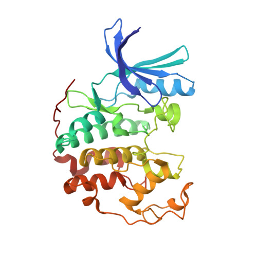
    5JQ5, 5JQ8

  • PubMed Abstract: 

    Deregulation of the cell cycle by mechanisms that lead to elevated activities of cyclin-dependent kinases (CDK) is a feature of many human diseases, cancer in particular. We identified small-molecule inhibitors that selectively inhibit CDK7, the kinase that phosphorylates cell-cycle CDKs to promote their activities. To investigate the selectivity of these inhibitors we used a combination of structural, biophysical, and modelling approaches. We determined the crystal structures of the CDK7-selective compounds ICEC0942 and ICEC0943 bound to CDK2, and used these to build models of inhibitor binding to CDK7. Molecular dynamics (MD) simulations of inhibitors bound to CDK2 and CDK7 generated possible models of inhibitor binding. To experimentally validate these models, we gathered isothermal titration calorimetry (ITC) binding data for recombinant wild-type and binding site mutants of CDK7 and CDK2. We identified specific residues of CDK7, notably Asp155, that are involved in determining inhibitor selectivity. Our MD simulations also show that the flexibility of the G-rich and activation loops of CDK7 is likely an important determinant of inhibitor specificity similar to CDK2.


  • Organizational Affiliation

    Section of Structural Biology, Department of Medicine, Imperial College London, South Kensington Campus, London, SW7 2AZ, UK.


Macromolecules
Find similar proteins by:  (by identity cutoff)  |  3D Structure
Entity ID: 1
MoleculeChains Sequence LengthOrganismDetailsImage
Cyclin-dependent kinase 2298Homo sapiensMutation(s): 0 
Gene Names: CDK2CDKN2
EC: 2.7.11.22
UniProt & NIH Common Fund Data Resources
Find proteins for P24941 (Homo sapiens)
Explore P24941 
Go to UniProtKB:  P24941
PHAROS:  P24941
GTEx:  ENSG00000123374 
Entity Groups  
Sequence Clusters30% Identity50% Identity70% Identity90% Identity95% Identity100% Identity
UniProt GroupP24941
Sequence Annotations
Expand
  • Reference Sequence
Small Molecules
Ligands 2 Unique
IDChains Name / Formula / InChI Key2D Diagram3D Interactions
I74
Query on I74

Download Ideal Coordinates CCD File 
C [auth A](3R,4R)-4-[[[7-[(phenylmethyl)amino]-3-propan-2-yl-pyrazolo[1,5-a]pyrimidin-5-yl]amino]methyl]piperidin-3-ol
C22 H30 N6 O
YCVGLKWJKIKVBI-MJGOQNOKSA-N
ACT
Query on ACT

Download Ideal Coordinates CCD File 
B [auth A]ACETATE ION
C2 H3 O2
QTBSBXVTEAMEQO-UHFFFAOYSA-M
Binding Affinity Annotations 
IDSourceBinding Affinity
I74 Binding MOAD:  5JQ5 Kd: 2341 (nM) from 1 assay(s)
Experimental Data & Validation

Experimental Data

  • Method: X-RAY DIFFRACTION
  • Resolution: 1.94 Å
  • R-Value Free: 0.241 
  • R-Value Work: 0.181 
  • R-Value Observed: 0.184 
  • Space Group: P 21 21 21
Unit Cell:
Length ( Å )Angle ( ˚ )
a = 53.65α = 90
b = 70.77β = 90
c = 72.27γ = 90
Software Package:
Software NamePurpose
REFMACrefinement
iMOSFLMdata reduction
SCALEPACKdata scaling
MOLREPphasing

Structure Validation

View Full Validation Report


Ligand Structure Quality Assessment 


Entry History 

Deposition Data

Revision History  (Full details and data files)

  • Version 1.0: 2017-02-08
    Type: Initial release
  • Version 1.1: 2017-02-15
    Changes: Database references
  • Version 1.2: 2017-03-15
    Changes: Database references
  • Version 1.3: 2024-01-10
    Changes: Data collection, Database references, Refinement description