5JON

Crystal structure of the unliganded form of HCN2 CNBD


Experimental Data Snapshot

  • Method: X-RAY DIFFRACTION
  • Resolution: 2.04 Å
  • R-Value Free: 0.221 
  • R-Value Work: 0.180 
  • R-Value Observed: 0.182 

wwPDB Validation   3D Report Full Report


This is version 2.1 of the entry. See complete history


Literature

Structure and dynamics underlying elementary ligand binding events in human pacemaking channels.

Goldschen-Ohm, M.P.Klenchin, V.A.White, D.S.Cowgill, J.B.Cui, Q.Goldsmith, R.H.Chanda, B.

(2016) Elife 5

  • DOI: https://doi.org/10.7554/eLife.20797
  • Primary Citation of Related Structures:  
    5JON

  • PubMed Abstract: 

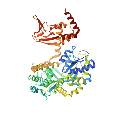
    Although molecular recognition is crucial for cellular signaling, mechanistic studies have relied primarily on ensemble measures that average over and thereby obscure underlying steps. Single-molecule observations that resolve these steps are lacking due to diffraction-limited resolution of single fluorophores at relevant concentrations. Here, we combined zero-mode waveguides with fluorescence resonance energy transfer (FRET) to directly observe binding at individual cyclic nucleotide-binding domains (CNBDs) from human pacemaker ion channels critical for heart and brain function. Our observations resolve the dynamics of multiple distinct steps underlying cyclic nucleotide regulation: a slow initial binding step that must select a 'receptive' conformation followed by a ligand-induced isomerization of the CNBD. X-ray structure of the apo CNBD and atomistic simulations reveal that the isomerization involves both local and global transitions. Our approach reveals fundamental mechanisms underpinning ligand regulation of pacemaker channels, and is generally applicable to weak-binding interactions governing a broad spectrum of signaling processes.


  • Organizational Affiliation

    Department of Neuroscience, University of Wisconsin-Madison, Madison, United States.


Macromolecules
Find similar proteins by:  (by identity cutoff)  |  3D Structure
Entity ID: 1
MoleculeChains Sequence LengthOrganismDetailsImage
Maltose-binding periplasmic protein,Potassium/sodium hyperpolarization-activated cyclic nucleotide-gated channel 2
A, B
517Escherichia coli O157:H7Mus musculus
This entity is chimeric
Mutation(s): 0 
Gene Names: malEZ5632ECs5017Hcn2Bcng2Hac1
UniProt
Find proteins for O88703 (Mus musculus)
Explore O88703 
Go to UniProtKB:  O88703
Find proteins for P0AEY0 (Escherichia coli O157:H7)
Explore P0AEY0 
Go to UniProtKB:  P0AEY0
Entity Groups  
Sequence Clusters30% Identity50% Identity70% Identity90% Identity95% Identity100% Identity
UniProt GroupsO88703P0AEY0
Sequence Annotations
Expand
  • Reference Sequence
Oligosaccharides

Help

Entity ID: 2
MoleculeChains Length2D Diagram Glycosylation3D Interactions
alpha-D-glucopyranose-(1-4)-alpha-D-glucopyranose
C, D
2N/A
Glycosylation Resources
GlyTouCan:  G07411ON
GlyCosmos:  G07411ON
Biologically Interesting Molecules (External Reference) 1 Unique
Experimental Data & Validation

Experimental Data

  • Method: X-RAY DIFFRACTION
  • Resolution: 2.04 Å
  • R-Value Free: 0.221 
  • R-Value Work: 0.180 
  • R-Value Observed: 0.182 
  • Space Group: P 1 21 1
Unit Cell:
Length ( Å )Angle ( ˚ )
a = 61.517α = 90
b = 42.01β = 90.85
c = 198.376γ = 90
Software Package:
Software NamePurpose
REFMACrefinement
HKL-2000data reduction
HKL-2000data scaling
PHASERphasing

Structure Validation

View Full Validation Report


Entry History & Funding Information

Deposition Data


Funding OrganizationLocationGrant Number
National Institutes of Health/National Institute of General Medical Sciences (NIH/NIGMS)United StatesR01-GM084140
National Institutes of Health/National Institute of Neurological Disorders and Stroke (NIH/NINDS)United StatesR01-NS081293

Revision History  (Full details and data files)

  • Version 1.0: 2016-11-30
    Type: Initial release
  • Version 1.1: 2016-12-21
    Changes: Database references
  • Version 1.2: 2017-09-06
    Changes: Author supporting evidence
  • Version 1.3: 2019-12-18
    Changes: Author supporting evidence
  • Version 2.0: 2020-07-29
    Type: Remediation
    Reason: Carbohydrate remediation
    Changes: Atomic model, Data collection, Derived calculations, Non-polymer description, Structure summary
  • Version 2.1: 2023-09-27
    Changes: Data collection, Database references, Refinement description, Structure summary