5JJM

Crystal Structure of Homodimeric Androgen Receptor Ligand-Binding Domain bound to DHT and LxxLL peptide


Experimental Data Snapshot

  • Method: X-RAY DIFFRACTION
  • Resolution: 2.15 Å
  • R-Value Free: 0.243 
  • R-Value Work: 0.201 
  • R-Value Observed: 0.203 

wwPDB Validation   3D Report Full Report


Ligand Structure Quality Assessment 


This is version 1.1 of the entry. See complete history


Literature

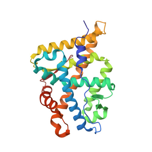
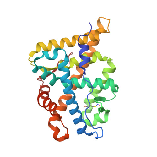
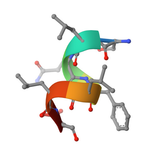
Structure of the homodimeric androgen receptor ligand-binding domain.

Nadal, M.Prekovic, S.Gallastegui, N.Helsen, C.Abella, M.Zielinska, K.Gay, M.Vilaseca, M.Taules, M.Houtsmuller, A.B.van Royen, M.E.Claessens, F.Fuentes-Prior, P.Estebanez-Perpina, E.

(2017) Nat Commun 8: 14388-14388

  • DOI: https://doi.org/10.1038/ncomms14388
  • Primary Citation of Related Structures:  
    5JJM

  • PubMed Abstract: 

    The androgen receptor (AR) plays a crucial role in normal physiology, development and metabolism as well as in the aetiology and treatment of diverse pathologies such as androgen insensitivity syndromes (AIS), male infertility and prostate cancer (PCa). Here we show that dimerization of AR ligand-binding domain (LBD) is induced by receptor agonists but not by antagonists. The 2.15-Å crystal structure of homodimeric, agonist- and coactivator peptide-bound AR-LBD unveils a 1,000-Å 2 large dimerization surface, which harbours over 40 previously unexplained AIS- and PCa-associated point mutations. An AIS mutation in the self-association interface (P767A) disrupts dimer formation in vivo, and has a detrimental effect on the transactivating properties of full-length AR, despite retained hormone-binding capacity. The conservation of essential residues suggests that the unveiled dimerization mechanism might be shared by other nuclear receptors. Our work defines AR-LBD homodimerization as an essential step in the proper functioning of this important transcription factor.


  • Organizational Affiliation

    Department of Biochemistry and Molecular Biomedicine, Institute of Biomedicine (IBUB) of the University of Barcelona (UB), Barcelona 08028, Spain.


Macromolecules
Find similar proteins by:  (by identity cutoff)  |  3D Structure
Entity ID: 1
MoleculeChains Sequence LengthOrganismDetailsImage
Androgen receptorA,
E [auth C],
G [auth D]
252Homo sapiensMutation(s): 0 
Gene Names: ARDHTRNR3C4
UniProt & NIH Common Fund Data Resources
Find proteins for P10275 (Homo sapiens)
Explore P10275 
Go to UniProtKB:  P10275
PHAROS:  P10275
GTEx:  ENSG00000169083 
Entity Groups  
Sequence Clusters30% Identity50% Identity70% Identity90% Identity95% Identity100% Identity
UniProt GroupP10275
Sequence Annotations
Expand
  • Reference Sequence

Find similar proteins by:  Sequence   |   3D Structure  

Entity ID: 2
MoleculeChains Sequence LengthOrganismDetailsImage
Unknown peptideB [auth M],
D [auth F],
F [auth K],
H [auth L]
3Homo sapiensMutation(s): 0 
Sequence Annotations
Expand
  • Reference Sequence
Find similar proteins by:  (by identity cutoff)  |  3D Structure
Entity ID: 3
MoleculeChains Sequence LengthOrganismDetailsImage
Androgen receptorC [auth B]252Homo sapiensMutation(s): 0 
Gene Names: ARDHTRNR3C4
UniProt & NIH Common Fund Data Resources
Find proteins for P10275 (Homo sapiens)
Explore P10275 
Go to UniProtKB:  P10275
PHAROS:  P10275
GTEx:  ENSG00000169083 
Entity Groups  
Sequence Clusters30% Identity50% Identity70% Identity90% Identity95% Identity100% Identity
UniProt GroupP10275
Sequence Annotations
Expand
  • Reference Sequence

Find similar proteins by:  Sequence   |   3D Structure  

Entity ID: 4
MoleculeChains Sequence LengthOrganismDetailsImage
uba3-derived peptideI [auth G],
J [auth H],
K [auth I],
L [auth J]
8Homo sapiensMutation(s): 0 
UniProt & NIH Common Fund Data Resources
Find proteins for Q8TBC4 (Homo sapiens)
Explore Q8TBC4 
Go to UniProtKB:  Q8TBC4
PHAROS:  Q8TBC4
GTEx:  ENSG00000144744 
Entity Groups  
UniProt GroupQ8TBC4
Sequence Annotations
Expand
  • Reference Sequence
Small Molecules
Ligands 8 Unique
IDChains Name / Formula / InChI Key2D Diagram3D Interactions
DHT
Query on DHT

Download Ideal Coordinates CCD File 
HA [auth D],
M [auth A],
S [auth B],
Z [auth C]
5-ALPHA-DIHYDROTESTOSTERONE
C19 H30 O2
NVKAWKQGWWIWPM-ABEVXSGRSA-N
PGE
Query on PGE

Download Ideal Coordinates CCD File 
DA [auth C]TRIETHYLENE GLYCOL
C6 H14 O4
ZIBGPFATKBEMQZ-UHFFFAOYSA-N
SO4
Query on SO4

Download Ideal Coordinates CCD File 
EA [auth C],
FA [auth C],
NA [auth D],
R [auth A],
Y [auth B]
SULFATE ION
O4 S
QAOWNCQODCNURD-UHFFFAOYSA-L
GOL
Query on GOL

Download Ideal Coordinates CCD File 
CA [auth C],
Q [auth A]
GLYCEROL
C3 H8 O3
PEDCQBHIVMGVHV-UHFFFAOYSA-N
ZN
Query on ZN

Download Ideal Coordinates CCD File 
AA [auth C],
GA [auth D],
N [auth A],
T [auth B]
ZINC ION
Zn
PTFCDOFLOPIGGS-UHFFFAOYSA-N
EDO
Query on EDO

Download Ideal Coordinates CCD File 
BA [auth C]
JA [auth D]
KA [auth D]
MA [auth D]
P [auth A]
BA [auth C],
JA [auth D],
KA [auth D],
MA [auth D],
P [auth A],
V [auth B],
W [auth B],
X [auth B]
1,2-ETHANEDIOL
C2 H6 O2
LYCAIKOWRPUZTN-UHFFFAOYSA-N
ACT
Query on ACT

Download Ideal Coordinates CCD File 
LA [auth D]ACETATE ION
C2 H3 O2
QTBSBXVTEAMEQO-UHFFFAOYSA-M
CL
Query on CL

Download Ideal Coordinates CCD File 
IA [auth D],
O [auth A],
U [auth B]
CHLORIDE ION
Cl
VEXZGXHMUGYJMC-UHFFFAOYSA-M
Modified Residues  2 Unique
IDChains TypeFormula2D DiagramParent
FME
Query on FME
B [auth M],
D [auth F],
F [auth K],
H [auth L]
L-PEPTIDE LINKINGC6 H11 N O3 SMET
SNC
Query on SNC
C [auth B]L-PEPTIDE LINKINGC3 H6 N2 O3 SCYS
Binding Affinity Annotations 
IDSourceBinding Affinity
DHT BindingDB:  5JJM Ki: min: 0.2, max: 10 (nM) from 12 assay(s)
IC50: min: 0.05, max: 18.5 (nM) from 14 assay(s)
EC50: min: 0.05, max: 20 (nM) from 14 assay(s)
Experimental Data & Validation

Experimental Data

  • Method: X-RAY DIFFRACTION
  • Resolution: 2.15 Å
  • R-Value Free: 0.243 
  • R-Value Work: 0.201 
  • R-Value Observed: 0.203 
  • Space Group: C 1 2 1
Unit Cell:
Length ( Å )Angle ( ˚ )
a = 91.09α = 90
b = 91.01β = 90.07
c = 157.23γ = 90
Software Package:
Software NamePurpose
REFMACrefinement
MOSFLMdata reduction
SCALAdata scaling
Cootmodel building

Structure Validation

View Full Validation Report


Ligand Structure Quality Assessment 


Entry History 

Deposition Data

Revision History  (Full details and data files)

  • Version 1.0: 2017-03-15
    Type: Initial release
  • Version 1.1: 2024-01-10
    Changes: Data collection, Database references, Derived calculations, Refinement description