5JIY

Structure of G9a SET-domain with Histone H3K9norLeucine mutant peptide and bound S-adenosylmethionine


Experimental Data Snapshot

  • Method: X-RAY DIFFRACTION
  • Resolution: 1.48 Å
  • R-Value Free: 0.233 
  • R-Value Work: 0.209 
  • R-Value Observed: 0.210 

wwPDB Validation   3D Report Full Report


Ligand Structure Quality Assessment 


This is version 1.2 of the entry. See complete history


Literature

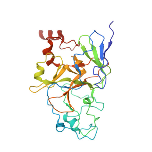
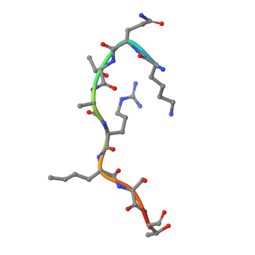
S-adenosyl methionine is necessary for inhibition of the methyltransferase G9a by the lysine 9 to methionine mutation on histone H3.

Jayaram, H.Hoelper, D.Jain, S.U.Cantone, N.Lundgren, S.M.Poy, F.Allis, C.D.Cummings, R.Bellon, S.Lewis, P.W.

(2016) Proc Natl Acad Sci U S A 113: 6182-6187

  • DOI: https://doi.org/10.1073/pnas.1605523113
  • Primary Citation of Related Structures:  
    5JHN, 5JIN, 5JIY, 5JJ0

  • PubMed Abstract: 

    Lysine to methionine (K-to-M) mutations in genes encoding histone H3 are thought to drive a subset of pediatric brain and bone cancers. These high-frequency K-to-M mutations occur at sites of methylation on histone H3, and tumors containing the mutant histones exhibit a global loss of specific histone methylation marks. Previous studies showed that K-to-M mutant histones, also known as oncohistones, are potent orthosteric inhibitors of specific Su(var)3-9, Enhancer-of-zeste, Trithorax (SET) domain methyltransferases. However, the biochemical and biophysical details of the interaction between K-to-M mutant histones and the respective SET domain methyltransferases are currently unknown. Here, we use the histone H3K9-directed methyltransferase G9a as a model to explore the mechanism of inhibition by K-to-M oncohistones. X-ray cocrystal structures revealed that the K9M residue of histone H3 occupies the active site cavity of G9a, and kinetic analysis indicates competitive inhibition of G9a by histone H3K9M. Additionally, we find that the cofactor S-adenosyl methionine (SAM) is necessary for stable interaction between G9a and H3K9M histone. Consistent with the formation of a ternary complex, we find that the inhibitory peptide is uncompetitive with regard to SAM. These data and others indicate that K-to-M oncohistones promote global loss of specific lysine methylation through sequestration and inhibition of SAM-bound SET domain methyltransferases.


  • Organizational Affiliation

    Constellation Pharmaceuticals, Cambridge, MA 02142;


Macromolecules
Find similar proteins by:  (by identity cutoff)  |  3D Structure
Entity ID: 1
MoleculeChains Sequence LengthOrganismDetailsImage
Histone-lysine N-methyltransferase EHMT2A,
C [auth B]
274Homo sapiensMutation(s): 0 
Gene Names: EHMT2BAT8C6orf30G9AKMT1CNG36
EC: 2.1.1 (PDB Primary Data), 2.1.1.43 (PDB Primary Data)
UniProt & NIH Common Fund Data Resources
Find proteins for Q96KQ7 (Homo sapiens)
Explore Q96KQ7 
Go to UniProtKB:  Q96KQ7
PHAROS:  Q96KQ7
GTEx:  ENSG00000204371 
Entity Groups  
Sequence Clusters30% Identity50% Identity70% Identity90% Identity95% Identity100% Identity
UniProt GroupQ96KQ7
Sequence Annotations
Expand
  • Reference Sequence

Find similar proteins by:  Sequence   |   3D Structure  

Entity ID: 2
MoleculeChains Sequence LengthOrganismDetailsImage
Histone H3.1 mutant peptide with H3K9nor-leucineB [auth F],
D [auth G]
11Homo sapiensMutation(s): 0 
UniProt & NIH Common Fund Data Resources
Find proteins for P68431 (Homo sapiens)
Explore P68431 
Go to UniProtKB:  P68431
PHAROS:  P68431
Entity Groups  
Sequence Clusters30% Identity50% Identity70% Identity90% Identity95% Identity100% Identity
UniProt GroupP68431
Sequence Annotations
Expand
  • Reference Sequence
Small Molecules
Ligands 2 Unique
IDChains Name / Formula / InChI Key2D Diagram3D Interactions
SAM
Query on SAM

Download Ideal Coordinates CCD File 
I [auth A],
N [auth B]
S-ADENOSYLMETHIONINE
C15 H22 N6 O5 S
MEFKEPWMEQBLKI-FCKMPRQPSA-N
ZN
Query on ZN

Download Ideal Coordinates CCD File 
E [auth A]
F [auth A]
G [auth A]
H [auth A]
J [auth B]
E [auth A],
F [auth A],
G [auth A],
H [auth A],
J [auth B],
K [auth B],
L [auth B],
M [auth B]
ZINC ION
Zn
PTFCDOFLOPIGGS-UHFFFAOYSA-N
Modified Residues  1 Unique
IDChains TypeFormula2D DiagramParent
NLE
Query on NLE
B [auth F],
D [auth G]
L-PEPTIDE LINKINGC6 H13 N O2LEU
Experimental Data & Validation

Experimental Data

  • Method: X-RAY DIFFRACTION
  • Resolution: 1.48 Å
  • R-Value Free: 0.233 
  • R-Value Work: 0.209 
  • R-Value Observed: 0.210 
  • Space Group: P 1 21 1
Unit Cell:
Length ( Å )Angle ( ˚ )
a = 56.611α = 90
b = 78.463β = 90.28
c = 73.084γ = 90
Software Package:
Software NamePurpose
REFMACrefinement
HKL-2000data reduction
SCALEPACKdata scaling
PHASERphasing

Structure Validation

View Full Validation Report


Ligand Structure Quality Assessment 


Entry History 

Deposition Data

Revision History  (Full details and data files)

  • Version 1.0: 2016-09-14
    Type: Initial release
  • Version 1.1: 2023-09-27
    Changes: Data collection, Database references, Derived calculations, Refinement description
  • Version 1.2: 2023-11-15
    Changes: Data collection