5JIC

Staphylococcus aureus Type II pantothenate kinase in complex with a pantothenate analog


Experimental Data Snapshot

  • Method: X-RAY DIFFRACTION
  • Resolution: 1.40 Å
  • R-Value Free: 0.174 
  • R-Value Work: 0.142 
  • R-Value Observed: 0.143 

wwPDB Validation   3D Report Full Report


Ligand Structure Quality Assessment 


This is version 1.2 of the entry. See complete history


Literature

Discovery of Potent Pantothenamide Inhibitors of Staphylococcus aureus Pantothenate Kinase through a Minimal SAR Study: Inhibition Is Due to Trapping of the Product.

Hughes, S.J.Barnard, L.Mottaghi, K.Tempel, W.Antoshchenko, T.Hong, B.S.Allali-Hassani, A.Smil, D.Vedadi, M.Strauss, E.Park, H.W.

(2016) ACS Infect Dis 2: 627-641

  • DOI: https://doi.org/10.1021/acsinfecdis.6b00090
  • Primary Citation of Related Structures:  
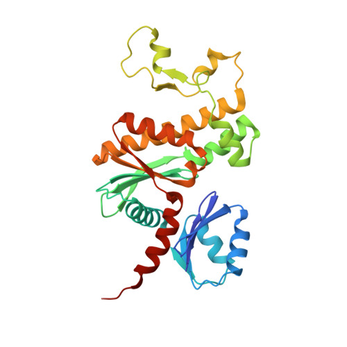
    4M7X, 4M7Y, 5ELZ, 5JIC

  • PubMed Abstract: 

    The potent antistaphylococcal activity of N-substituted pantothenamides (PanAms) has been shown to at least partially be due to the inhibition of Staphylococcus aureus's atypical type II pantothenate kinase (SaPanK II ), the first enzyme of coenzyme A biosynthesis. This mechanism of action follows from SaPanK II having a binding mode for PanAms that is distinct from those of other PanKs. To dissect the molecular interactions responsible for PanAm inhibitory activity, we conducted a mini SAR study in tandem with the cocrystallization of SaPanK II with two classic PanAms (N5-Pan and N7-Pan), culminating in the synthesis and characterization of two new PanAms, N-Pip-PanAm and MeO-N5-PanAm. The cocrystal structures showed that all of the PanAms are phosphorylated by SaPanK II but remain bound at the active site; this occurs primarily through interactions with Tyr240' and Thr172'. Kinetic analysis showed a strong correlation between k cat (slow PanAm turnover) and IC 50 (inhibition of pantothenate phosphorylation) values, suggesting that SaPanK II inhibition occurs via a delay in product release. In-depth analysis of the PanAm-bound structures showed that the capacity for accepting a hydrogen bond from the amide of Thr172' was a stronger determinant for PanAm potency than the capacity to π-stack with Tyr240'. The two new PanAms, N-Pip-PanAm and MeO-N5-PanAm, effectively combine both hydrogen bonding and hydrophobic interactions, resulting in the most potent SaPanK II inhibition described to date. Taken together, our results are consistent with an inhibition mechanism wherein PanAms act as SaPanK II substrates that remain bound upon phosphorylation. The phospho-PanAm-SaPanK II interactions described herein may help future antistaphylococcal drug development.


  • Organizational Affiliation

    Department of Biochemistry, Stellenbosch University , Stellenbosch 7600, South Africa.


Macromolecules
Find similar proteins by:  (by identity cutoff)  |  3D Structure
Entity ID: 1
MoleculeChains Sequence LengthOrganismDetailsImage
Type II pantothenate kinase273Staphylococcus aureus subsp. aureus MW2Mutation(s): 0 
Gene Names: coaWMW2054
EC: 2.7.1.33
UniProt
Find proteins for Q2FWC7 (Staphylococcus aureus (strain NCTC 8325 / PS 47))
Explore Q2FWC7 
Go to UniProtKB:  Q2FWC7
Entity Groups  
Sequence Clusters30% Identity50% Identity70% Identity90% Identity95% Identity100% Identity
UniProt GroupQ2FWC7
Sequence Annotations
Expand
  • Reference Sequence
Small Molecules
Ligands 3 Unique
IDChains Name / Formula / InChI Key2D Diagram3D Interactions
ADP
Query on ADP

Download Ideal Coordinates CCD File 
C [auth A]ADENOSINE-5'-DIPHOSPHATE
C10 H15 N5 O10 P2
XTWYTFMLZFPYCI-KQYNXXCUSA-N
N7E
Query on N7E

Download Ideal Coordinates CCD File 
D [auth A]N~3~-[(2R)-2-hydroxy-3,3-dimethyl-4-(phosphonooxy)butanoyl]-N-(5-methoxypentyl)-beta-alaninamide
C15 H31 N2 O8 P
FOBYDOXAQNKICH-ZDUSSCGKSA-N
MG
Query on MG

Download Ideal Coordinates CCD File 
B [auth A]MAGNESIUM ION
Mg
JLVVSXFLKOJNIY-UHFFFAOYSA-N
Binding Affinity Annotations 
IDSourceBinding Affinity
N7E Binding MOAD:  5JIC IC50: 130 (nM) from 1 assay(s)
Experimental Data & Validation

Experimental Data

  • Method: X-RAY DIFFRACTION
  • Resolution: 1.40 Å
  • R-Value Free: 0.174 
  • R-Value Work: 0.142 
  • R-Value Observed: 0.143 
  • Space Group: C 2 2 21
Unit Cell:
Length ( Å )Angle ( ˚ )
a = 57.387α = 90
b = 136.746β = 90
c = 73.947γ = 90
Software Package:
Software NamePurpose
XDSdata reduction
SCALAdata scaling
MOLREPphasing
REFMACrefinement
PDB_EXTRACTdata extraction

Structure Validation

View Full Validation Report


Ligand Structure Quality Assessment 


Entry History 

Deposition Data

Revision History  (Full details and data files)

  • Version 1.0: 2016-08-10
    Type: Initial release
  • Version 1.1: 2016-11-02
    Changes: Database references
  • Version 1.2: 2023-09-27
    Changes: Data collection, Database references, Derived calculations, Refinement description