A comparative structural analysis reveals distinctive features of co-factor binding and substrate specificity in plant aldo-keto reductases.
Giuseppe, P.O., Santos, M.L., Sousa, S.M., Koch, K.E., Yunes, J.A., Aparicio, R., Murakami, M.T.(2016) Biochem Biophys Res Commun 474: 696-701
- PubMed: 27154221
- DOI: https://doi.org/10.1016/j.bbrc.2016.05.011
- Primary Citation of Related Structures:
5JH1, 5JH2 - PubMed Abstract:
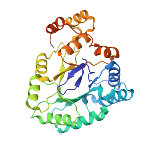
Plant aldo-keto reductases of the AKR4C subfamily play key roles during stress and are attractive targets for developing stress-tolerant crops. However, these AKR4Cs show little to no activity with previously-envisioned sugar substrates. We hypothesized a structural basis for the distinctive cofactor binding and substrate specificity of these plant enzymes. To test this, we solved the crystal structure of a novel AKR4C subfamily member, the AKR4C7 from maize, in the apo form and in complex with NADP(+). The binary complex revealed an intermediate state of cofactor binding that preceded closure of Loop B, and also indicated that conformational changes upon substrate binding are required to induce a catalytically-favorable conformation of the active-site pocket. Comparative structural analyses of homologues (AKR1B1, AKR4C8 and AKR4C9) showed that evolutionary redesign of plant AKR4Cs weakened interactions that stabilize the closed conformation of Loop B. This in turn decreased cofactor affinity and altered configuration of the substrate-binding site. We propose that these structural modifications contribute to impairment of sugar reductase activity in favor of other substrates in the plant AKR4C subgroup, and that catalysis involves a three-step process relevant to other AKRs.
Organizational Affiliation:
Biosciences National Laboratory (LNBio), National Center for Research in Energy and Materials (CNPEM), Campinas, SP, Brazil.