5JBT

Mesotrypsin in complex with cleaved amyloid precursor like protein 2 inhibitor (APLP2)


Experimental Data Snapshot

  • Method: X-RAY DIFFRACTION
  • Resolution: 1.40 Å
  • R-Value Free: 0.199 
  • R-Value Work: 0.163 
  • R-Value Observed: 0.165 

wwPDB Validation   3D Report Full Report


This is version 1.5 of the entry. See complete history


Literature

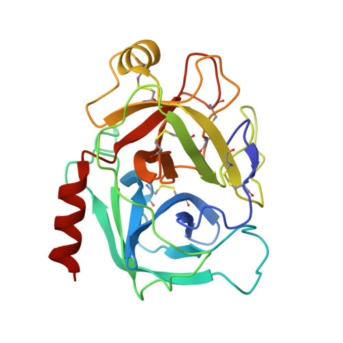
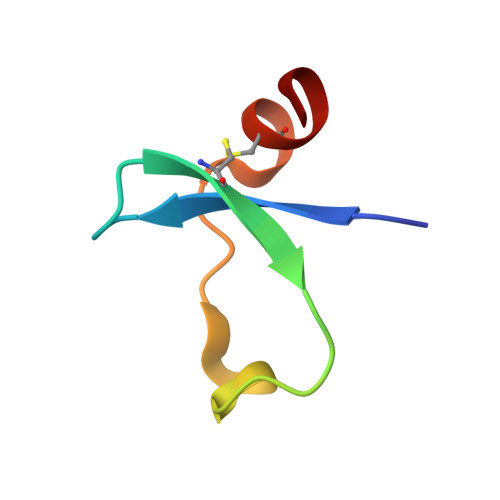
An Acrobatic Substrate Metamorphosis Reveals a Requirement for Substrate Conformational Dynamics in Trypsin Proteolysis.

Kayode, O.Wang, R.Pendlebury, D.F.Cohen, I.Henin, R.D.Hockla, A.Soares, A.S.Papo, N.Caulfield, T.R.Radisky, E.S.

(2016) J Biol Chem 291: 26304-26319

  • DOI: https://doi.org/10.1074/jbc.M116.758417
  • Primary Citation of Related Structures:  
    5JBT

  • PubMed Abstract: 

    The molecular basis of enzyme catalytic power and specificity derives from dynamic interactions between enzyme and substrate during catalysis. Although considerable effort has been devoted to understanding how conformational dynamics within enzymes affect catalysis, the role of conformational dynamics within protein substrates has not been addressed. Here, we examine the importance of substrate dynamics in the cleavage of Kunitz-bovine pancreatic trypsin inhibitor protease inhibitors by mesotrypsin, finding that the varied conformational dynamics of structurally similar substrates can profoundly impact the rate of catalysis. A 1.4-Å crystal structure of a mesotrypsin-product complex formed with a rapidly cleaved substrate reveals a dramatic conformational change in the substrate upon proteolysis. By using long all-atom molecular dynamics simulations of acyl-enzyme intermediates with proteolysis rates spanning 3 orders of magnitude, we identify global and local dynamic features of substrates on the nanosecond-microsecond time scale that correlate with enzymatic rates and explain differential susceptibility to proteolysis. By integrating multiple enhanced sampling methods for molecular dynamics, we model a viable conformational pathway between substrate-like and product-like states, linking substrate dynamics on the nanosecond-microsecond time scale with large collective substrate motions on the much slower time scale of catalysis. Our findings implicate substrate flexibility as a critical determinant of catalysis.


  • Organizational Affiliation

    From the Departments of Cancer Biology and.


Macromolecules
Find similar proteins by:  (by identity cutoff)  |  3D Structure
Entity ID: 1
MoleculeChains Sequence LengthOrganismDetailsImage
PRSS3 protein224Homo sapiensMutation(s): 1 
Gene Names: PRSS3
UniProt & NIH Common Fund Data Resources
Find proteins for P35030 (Homo sapiens)
Explore P35030 
Go to UniProtKB:  P35030
PHAROS:  P35030
GTEx:  ENSG00000010438 
Entity Groups  
Sequence Clusters30% Identity50% Identity70% Identity90% Identity95% Identity100% Identity
UniProt GroupP35030
Sequence Annotations
Expand
  • Reference Sequence

Find similar proteins by:  Sequence   |   3D Structure  

Entity ID: 2
MoleculeChains Sequence LengthOrganismDetailsImage
Amyloid-like protein 2B [auth X]14Homo sapiensMutation(s): 0 
Gene Names: APLP2APPL2
UniProt & NIH Common Fund Data Resources
Find proteins for Q06481 (Homo sapiens)
Explore Q06481 
Go to UniProtKB:  Q06481
PHAROS:  Q06481
GTEx:  ENSG00000084234 
Entity Groups  
Sequence Clusters30% Identity50% Identity70% Identity90% Identity95% Identity100% Identity
UniProt GroupQ06481
Sequence Annotations
Expand
  • Reference Sequence
Find similar proteins by:  (by identity cutoff)  |  3D Structure
Entity ID: 3
MoleculeChains Sequence LengthOrganismDetailsImage
Amyloid-like protein 2C [auth Y]38Homo sapiensMutation(s): 0 
Gene Names: APLP2APPL2
UniProt & NIH Common Fund Data Resources
Find proteins for Q06481 (Homo sapiens)
Explore Q06481 
Go to UniProtKB:  Q06481
PHAROS:  Q06481
GTEx:  ENSG00000084234 
Entity Groups  
Sequence Clusters30% Identity50% Identity70% Identity90% Identity95% Identity100% Identity
UniProt GroupQ06481
Sequence Annotations
Expand
  • Reference Sequence
Experimental Data & Validation

Experimental Data

  • Method: X-RAY DIFFRACTION
  • Resolution: 1.40 Å
  • R-Value Free: 0.199 
  • R-Value Work: 0.163 
  • R-Value Observed: 0.165 
  • Space Group: P 21 21 2
Unit Cell:
Length ( Å )Angle ( ˚ )
a = 98.956α = 90
b = 54.539β = 90
c = 56.632γ = 90
Software Package:
Software NamePurpose
REFMACrefinement
HKL-2000data scaling
PDB_EXTRACTdata extraction
HKL-2000data reduction

Structure Validation

View Full Validation Report


Entry History & Funding Information

Deposition Data


Funding OrganizationLocationGrant Number
National Institutes of Health/National Cancer Institute (NIH/NCI)United StatesR01CA154387

Revision History  (Full details and data files)

  • Version 1.0: 2016-11-09
    Type: Initial release
  • Version 1.1: 2016-11-16
    Changes: Refinement description
  • Version 1.2: 2016-12-28
    Changes: Database references
  • Version 1.3: 2017-09-06
    Changes: Author supporting evidence
  • Version 1.4: 2019-12-04
    Changes: Author supporting evidence
  • Version 1.5: 2023-09-27
    Changes: Data collection, Database references, Refinement description