5JBN

Crystal Structure of Apo Phosphopantetheine Adenylyltransferase (PPAT/CoaD) from E. coli


Experimental Data Snapshot

  • Method: X-RAY DIFFRACTION
  • Resolution: 1.45 Å
  • R-Value Free: 0.182 
  • R-Value Work: 0.145 
  • R-Value Observed: 0.147 

wwPDB Validation   3D Report Full Report


This is version 1.2 of the entry. See complete history


Literature

Facilitating unambiguous NMR assignments and enabling higher probe density through selective labeling of all methyl containing amino acids.

Proudfoot, A.Frank, A.O.Ruggiu, F.Mamo, M.Lingel, A.

(2016) J Biomol NMR 65: 15-27

  • DOI: https://doi.org/10.1007/s10858-016-0032-2
  • Primary Citation of Related Structures:  
    5JBN

  • PubMed Abstract: 

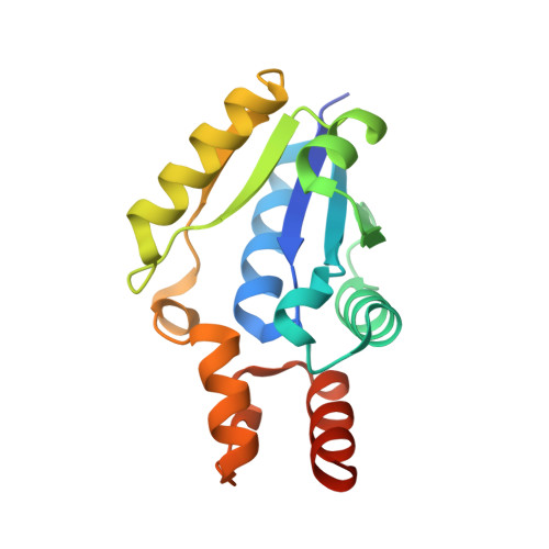
    The deuteration of proteins and selective labeling of side chain methyl groups has greatly enhanced the molecular weight range of proteins and protein complexes which can be studied using solution NMR spectroscopy. Protocols for the selective labeling of all six methyl group containing amino acids individually are available, however to date, only a maximum of five amino acids have been labeled simultaneously. Here, we describe a new methodology for the simultaneous, selective labeling of all six methyl containing amino acids using the 115 kDa homohexameric enzyme CoaD from E. coli as a model system. The utility of the labeling protocol is demonstrated by efficiently and unambiguously assigning all methyl groups in the enzymatic active site using a single 4D (13)C-resolved HMQC-NOESY-HMQC experiment, in conjunction with a crystal structure. Furthermore, the six fold labeled protein was employed to characterize the interaction between the substrate analogue (R)-pantetheine and CoaD by chemical shift perturbations, demonstrating the benefit of the increased probe density.


  • Organizational Affiliation

    Novartis Institutes for BioMedical Research, 5300 Chiron Way, Emeryville, CA, 94608, USA.


Macromolecules
Find similar proteins by:  (by identity cutoff)  |  3D Structure
Entity ID: 1
MoleculeChains Sequence LengthOrganismDetailsImage
Phosphopantetheine adenylyltransferase
A, B
170Escherichia coli K-12Mutation(s): 0 
Gene Names: coaDkdtByicAb3634JW3609
EC: 2.7.7.3
UniProt
Find proteins for P0A6I6 (Escherichia coli (strain K12))
Explore P0A6I6 
Go to UniProtKB:  P0A6I6
Entity Groups  
Sequence Clusters30% Identity50% Identity70% Identity90% Identity95% Identity100% Identity
UniProt GroupP0A6I6
Sequence Annotations
Expand
  • Reference Sequence
Experimental Data & Validation

Experimental Data

  • Method: X-RAY DIFFRACTION
  • Resolution: 1.45 Å
  • R-Value Free: 0.182 
  • R-Value Work: 0.145 
  • R-Value Observed: 0.147 
  • Space Group: I 2 3
Unit Cell:
Length ( Å )Angle ( ˚ )
a = 136.176α = 90
b = 136.176β = 90
c = 136.176γ = 90
Software Package:
Software NamePurpose
PHENIXrefinement
XDSdata reduction
SCALAdata scaling

Structure Validation

View Full Validation Report


Entry History 

Deposition Data

Revision History  (Full details and data files)

  • Version 1.0: 2016-05-25
    Type: Initial release
  • Version 1.1: 2016-07-06
    Changes: Database references
  • Version 1.2: 2024-03-06
    Changes: Data collection, Database references, Derived calculations