


Crystal structure of a putative alpha-ketoglutarate dependent 2,4-D dioxygenase from Burkholderia xenovorans
Conrady, D.G., Dranow, D.M., Lorimer, D., Edwards, T.E.To be published.
Experimental Data Snapshot
wwPDB Validation 3D Report Full Report

Entity ID: 1 | |||||
|---|---|---|---|---|---|
| Molecule | Chains | Sequence Length | Organism | Details | Image |
| Putative alpha KG dependent 2,4-D dioxygenase | 309 | Paraburkholderia xenovorans LB400 | Mutation(s): 0 Gene Names: Bxe_B2152 EC: 1.14.11 | 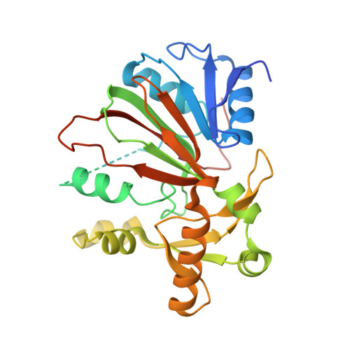 | |
UniProt | |||||
Find proteins for Q13Q05 (Paraburkholderia xenovorans (strain LB400)) Explore Q13Q05 Go to UniProtKB: Q13Q05 | |||||
Entity Groups | |||||
| Sequence Clusters | 30% Identity50% Identity70% Identity90% Identity95% Identity100% Identity | ||||
| UniProt Group | Q13Q05 | ||||
Sequence AnnotationsExpand | |||||
| |||||
| Ligands 2 Unique | |||||
|---|---|---|---|---|---|
| ID | Chains | Name / Formula / InChI Key | 2D Diagram | 3D Interactions | |
| ACT Query on ACT | H [auth C] | ACETATE ION C2 H3 O2 QTBSBXVTEAMEQO-UHFFFAOYSA-M |  | ||
| FE2 Query on FE2 | E [auth A], F [auth B], G [auth C], I [auth D] | FE (II) ION Fe CWYNVVGOOAEACU-UHFFFAOYSA-N |  | ||
| Length ( Å ) | Angle ( ˚ ) |
|---|---|
| a = 84.7 | α = 90 |
| b = 77.88 | β = 92.11 |
| c = 92.14 | γ = 90 |
| Software Name | Purpose |
|---|---|
| XSCALE | data scaling |
| MOLREP | phasing |
| PHENIX | refinement |
| PDB_EXTRACT | data extraction |
| XDS | data reduction |
RCSB PDB (citation) is hosted by
RCSB PDB is a member of the

