


The structure of Synechococcus elongatus enolase reveals key aspects of phosphoenolpyruvate binding
Gonzalez, J.M., Marti-Arbona, R., Chen, J.C.-H., Unkefer, C.J.(2022) Acta Crystallogr F Struct Biol Commun F78: 177-184
Experimental Data Snapshot
(2022) Acta Crystallogr F Struct Biol Commun F78: 177-184
Entity ID: 1 | |||||
|---|---|---|---|---|---|
| Molecule | Chains | Sequence Length | Organism | Details | Image |
| Enolase | 424 | Synechococcus elongatus PCC 7942 = FACHB-805 | Mutation(s): 0 Gene Names: eno, Synpcc7942_0639 EC: 4.2.1.11 | 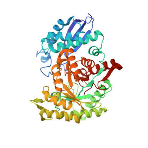 | |
UniProt | |||||
Find proteins for Q5N3P4 (Synechococcus sp. (strain ATCC 27144 / PCC 6301 / SAUG 1402/1)) Explore Q5N3P4 Go to UniProtKB: Q5N3P4 | |||||
Entity Groups | |||||
| Sequence Clusters | 30% Identity50% Identity70% Identity90% Identity95% Identity100% Identity | ||||
| UniProt Group | Q5N3P4 | ||||
Sequence AnnotationsExpand | |||||
| |||||
| Ligands 3 Unique | |||||
|---|---|---|---|---|---|
| ID | Chains | Name / Formula / InChI Key | 2D Diagram | 3D Interactions | |
| PEP (Subject of Investigation/LOI) Query on PEP | C [auth A], I [auth B] | PHOSPHOENOLPYRUVATE C3 H5 O6 P DTBNBXWJWCWCIK-UHFFFAOYSA-N |  | ||
| ACT Query on ACT | H [auth A], M [auth B] | ACETATE ION C2 H3 O2 QTBSBXVTEAMEQO-UHFFFAOYSA-M |  | ||
| CA (Subject of Investigation/LOI) Query on CA | D [auth A] E [auth A] F [auth A] G [auth A] J [auth B] | CALCIUM ION Ca BHPQYMZQTOCNFJ-UHFFFAOYSA-N |  | ||
| Length ( Å ) | Angle ( ˚ ) |
|---|---|
| a = 164.23 | α = 90 |
| b = 164.23 | β = 90 |
| c = 72.68 | γ = 90 |
| Software Name | Purpose |
|---|---|
| REFMAC | refinement |
| Aimless | data scaling |
| PDB_EXTRACT | data extraction |
| iMOSFLM | data reduction |
| PHASER | phasing |
| Funding Organization | Location | Grant Number |
|---|---|---|
| National Scientific and Technical Research Council (CONICET) | Argentina | PIP0024 |
RCSB PDB (citation) is hosted by
RCSB PDB is a member of the

