5IXE

1.75A RESOLUTION STRUCTURE OF 5-Fluoroindole BOUND BETA-GLYCOSIDASE (W33G) FROM SULFOLOBUS SOLFATARICUS


Experimental Data Snapshot

  • Method: X-RAY DIFFRACTION
  • Resolution: 1.75 Å
  • R-Value Free: 0.173 
  • R-Value Work: 0.153 
  • R-Value Observed: 0.154 

wwPDB Validation   3D Report Full Report


This is version 1.4 of the entry. See complete history


Literature

Full and Partial Agonism of a Designed Enzyme Switch.

Budiardjo, S.J.Licknack, T.J.Cory, M.B.Kapros, D.Roy, A.Lovell, S.Douglas, J.Karanicolas, J.

(2016) ACS Synth Biol 5: 1475-1484

  • DOI: https://doi.org/10.1021/acssynbio.6b00097
  • Primary Citation of Related Structures:  
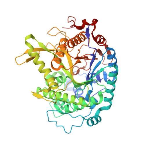
    5IXE

  • PubMed Abstract: 

    Chemical biology has long sought to build protein switches for use in molecular diagnostics, imaging, and synthetic biology. The overarching challenge for any type of engineered protein switch is the ability to respond in a selective and predictable manner that caters to the specific environments and time scales needed for the application at hand. We previously described a general method to design switchable proteins, called "chemical rescue of structure", that builds de novo allosteric control sites directly into a protein's functional domain. This approach entails first carving out a buried cavity in a protein via mutation, such that the protein's structure is disrupted and activity is lost. An exogenous ligand is subsequently added to substitute for the atoms that were removed by mutation, restoring the protein's structure and thus its activity. Here, we begin to ask what principles dictate such switches' response to different activating ligands. Using a redesigned β-glycosidase enzyme as our model system, we find that the designed effector site is quite malleable and can accommodate both larger and smaller ligands, but that optimal rescue comes only from a ligand that perfectly replaces the deleted atoms. Guided by these principles, we then altered the shape of this cavity by using different cavity-forming mutations, and predicted different ligands that would better complement these new cavities. These findings demonstrate how the protein switch's response can be tuned via small changes to the ligand with respect to the binding cavity, and ultimately enabled us to design an improved switch. We anticipate that these insights will help enable the design of future systems that tune other aspects of protein activity, whereby, like evolved protein receptors, remolding the effector site can also adjust additional outputs such as substrate selectivity and activation of downstream signaling pathways.


  • Organizational Affiliation

    Center for Computational Biology, ‡Department of Molecular Biosciences, §High Throughput Screening Laboratory, ∥Protein Structure Laboratory, ⊥Molecular Structures Group The University of Kansas , 2030 Becker Drive, Lawrence, Kansas 66045-7534, United States.


Macromolecules
Find similar proteins by:  (by identity cutoff)  |  3D Structure
Entity ID: 1
MoleculeChains Sequence LengthOrganismDetailsImage
Beta-galactosidase
A, B
489Saccharolobus solfataricus P2Mutation(s): 1 
Gene Names: lacSSSO3019
EC: 3.2.1.23
UniProt
Find proteins for P22498 (Saccharolobus solfataricus (strain ATCC 35092 / DSM 1617 / JCM 11322 / P2))
Explore P22498 
Go to UniProtKB:  P22498
Entity Groups  
Sequence Clusters30% Identity50% Identity70% Identity90% Identity95% Identity100% Identity
UniProt GroupP22498
Sequence Annotations
Expand
  • Reference Sequence
Small Molecules
Ligands 3 Unique
IDChains Name / Formula / InChI Key2D Diagram3D Interactions
14O
Query on 14O

Download Ideal Coordinates CCD File 
C [auth A],
H [auth B]
5-fluoro-1H-indole
C8 H6 F N
ODFFPRGJZRXNHZ-UHFFFAOYSA-N
MPD
Query on MPD

Download Ideal Coordinates CCD File 
D [auth A](4S)-2-METHYL-2,4-PENTANEDIOL
C6 H14 O2
SVTBMSDMJJWYQN-YFKPBYRVSA-N
CL
Query on CL

Download Ideal Coordinates CCD File 
E [auth A]
F [auth A]
G [auth A]
I [auth B]
J [auth B]
E [auth A],
F [auth A],
G [auth A],
I [auth B],
J [auth B],
K [auth B]
CHLORIDE ION
Cl
VEXZGXHMUGYJMC-UHFFFAOYSA-M
Binding Affinity Annotations 
IDSourceBinding Affinity
14O Binding MOAD:  5IXE Kd: 2.30e+6 (nM) from 1 assay(s)
Experimental Data & Validation

Experimental Data

  • Method: X-RAY DIFFRACTION
  • Resolution: 1.75 Å
  • R-Value Free: 0.173 
  • R-Value Work: 0.153 
  • R-Value Observed: 0.154 
  • Space Group: P 31 2 1
Unit Cell:
Length ( Å )Angle ( ˚ )
a = 167.706α = 90
b = 167.706β = 90
c = 95.871γ = 120
Software Package:
Software NamePurpose
Aimlessdata scaling
PHASERphasing
PHENIXrefinement
PDB_EXTRACTdata extraction
XDSdata reduction

Structure Validation

View Full Validation Report


Entry History & Funding Information

Deposition Data


Funding OrganizationLocationGrant Number
National Science Foundation (NSF, United States)United StatesMCB130049

Revision History  (Full details and data files)

  • Version 1.0: 2016-07-20
    Type: Initial release
  • Version 1.1: 2016-12-28
    Changes: Database references
  • Version 1.2: 2017-09-20
    Changes: Author supporting evidence
  • Version 1.3: 2019-11-27
    Changes: Author supporting evidence, Database references
  • Version 1.4: 2023-09-27
    Changes: Data collection, Database references, Refinement description