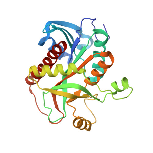
Crystal structure of Escherichia coli purine nucleoside phosphorylase in complex with 7-deazahypoxanthine.
Timofeev, V.I., Zhukhlistova, N.E., Abramchik, Y.A., Fateev, I.I., Kostromina, M.A., Muravieva, T.I., Esipov, R.S., Kuranova, I.P.(2018) Acta Crystallogr F Struct Biol Commun 74: 355-362
- PubMed: 29870020
- DOI: https://doi.org/10.1107/S2053230X18006337
- Primary Citation of Related Structures:
5IU6 - PubMed Abstract:
Purine nucleoside phosphorylases (EC 2.4.2.1; PNPs) reversibly catalyze the phosphorolytic cleavage of glycosidic bonds in purine nucleosides to generate ribose 1-phosphate and a free purine base, and are key enzymes in the salvage pathway of purine biosynthesis. They also catalyze the transfer of pentosyl groups between purine bases (the transglycosylation reaction) and are widely used for the synthesis of biologically important analogues of natural nucleosides, including a number of anticancer and antiviral drugs. Potent inhibitors of PNPs are used in chemotherapeutic applications. The detailed study of the binding of purine bases and their derivatives in the active site of PNPs is of particular interest in order to understand the mechanism of enzyme action and for the development of new enzyme inhibitors. Here, it is shown that 7-deazahypoxanthine (7DHX) is a noncompetitive inhibitor of the phosphorolysis of inosine by recombinant Escherichia coli PNP (EcPNP) with an inhibition constant K i of 0.13 mM. A crystal of EcPNP in complex with 7DHX was obtained in microgravity by the counter-diffusion technique and the three-dimensional structure of the EcPNP-7DHX complex was solved by molecular replacement at 2.51 Å resolution using an X-ray data set collected at the SPring-8 synchrotron-radiation facility, Japan. The crystals belonged to space group P6 1 22, with unit-cell parameters a = b = 120.370, c = 238.971 Å, and contained three subunits of the hexameric enzyme molecule in the asymmetric unit. The 7DHX molecule was located with full occupancy in the active site of each of the three crystallographically independent enzyme subunits. The position of 7DHX overlapped with the positions occupied by purine bases in similar PNP complexes. However, the orientation of the 7DHX molecule differs from those of other bases: it is rotated by ∼180° relative to other bases. The peculiarities of the arrangement of 7DHX in the EcPNP active site are discussed.
Organizational Affiliation:
Shubnikov Institute of Crystallography, Federal Scientific Research Centre `Crystallography and Photonics' of Russian Academy of Sciences, Leninsky Prospekt 59, Moscow 119333, Russian Federation.