5ITV

Crystal structure of Bacillus subtilis BacC Dihydroanticapsin 7-dehydrogenase in complex with NADH


Experimental Data Snapshot

  • Method: X-RAY DIFFRACTION
  • Resolution: 2.26 Å
  • R-Value Free: 0.221 
  • R-Value Work: 0.207 
  • R-Value Observed: 0.208 

wwPDB Validation   3D Report Full Report


Ligand Structure Quality Assessment 


This is version 1.2 of the entry. See complete history


Literature

Probing the influence of non-covalent contact networks identified by charge density analysis on the oxidoreductase BacC.

Perinbam, K.Balaram, H.Guru Row, T.N.Gopal, B.

(2017) Protein Eng Des Sel 30: 265-272

  • DOI: https://doi.org/10.1093/protein/gzx006
  • Primary Citation of Related Structures:  
    5ITV, 5ITW

  • PubMed Abstract: 

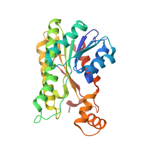
    Bacillus subtilis BacC is an oxidoreductase involved in the biosynthesis of the potent antibiotic bacilysin. The crystal structure of BacC was determined at 1.19 Å resolution. An experimental charge density approach was used to calculate non-covalent interactions within the monomer and across the dimeric interface of BacC. This interaction network, in turn, enabled an analysis of non-covalently connected paths that span the protein structure. One of the pathways of non-covalent interactions was examined by mutational analysis. Biochemical analysis of BacC mutants with potential disruptions in non-covalent interactions along this path revealed that residues that form nodes in pathways of non-covalent interactions influence catalytic activity more than others in a similar chemical environment. Furthermore, we note that nodes in the non-covalent interaction networks are co-localized with compensatory mutation sites identified by multiple sequence alignment of proteins with low sequence similarity to BacC. Put together, this analysis supports the hypothesis that non-covalent nodes represent conserved structural features that can impact the catalytic activity of an enzyme.


  • Organizational Affiliation

    Molecular Biophysics Unit, Indian Institute of Science, Bangalore 560012, India.


Macromolecules
Find similar proteins by:  (by identity cutoff)  |  3D Structure
Entity ID: 1
MoleculeChains Sequence LengthOrganismDetailsImage
Dihydroanticapsin 7-dehydrogenase
A, B, C, D
255Bacillus subtilis subsp. subtilis str. 168Mutation(s): 0 
Gene Names: bacCywfDBSU37720ipa-82d
EC: 1.1.1.385
UniProt
Find proteins for P39640 (Bacillus subtilis (strain 168))
Explore P39640 
Go to UniProtKB:  P39640
Entity Groups  
Sequence Clusters30% Identity50% Identity70% Identity90% Identity95% Identity100% Identity
UniProt GroupP39640
Sequence Annotations
Expand
  • Reference Sequence
Experimental Data & Validation

Experimental Data

  • Method: X-RAY DIFFRACTION
  • Resolution: 2.26 Å
  • R-Value Free: 0.221 
  • R-Value Work: 0.207 
  • R-Value Observed: 0.208 
  • Space Group: P 1 21 1
Unit Cell:
Length ( Å )Angle ( ˚ )
a = 74.75α = 90
b = 88.08β = 106.44
c = 78.61γ = 90
Software Package:
Software NamePurpose
REFMACrefinement
iMOSFLMdata reduction
SCALAdata scaling
PHASERphasing

Structure Validation

View Full Validation Report


Ligand Structure Quality Assessment 


Entry History 

Deposition Data

Revision History  (Full details and data files)

  • Version 1.0: 2017-02-22
    Type: Initial release
  • Version 1.1: 2017-12-06
    Changes: Database references
  • Version 1.2: 2024-03-20
    Changes: Data collection, Database references, Refinement description