


Crystal Structures of Dehydratase Domains from trans-AT Polyketide Biosynthetic Pathways
Jakob, R.P., Muller, R., Herbst, D.A., Maier, T.To be published.
Experimental Data Snapshot
wwPDB Validation 3D Report Full Report

Entity ID: 1 | |||||
|---|---|---|---|---|---|
| Molecule | Chains | Sequence Length | Organism | Details | Image |
| MlnD | 312 | Bacillus velezensis FZB42 | Mutation(s): 0 Gene Names: mlnD, RBAM_014360 | 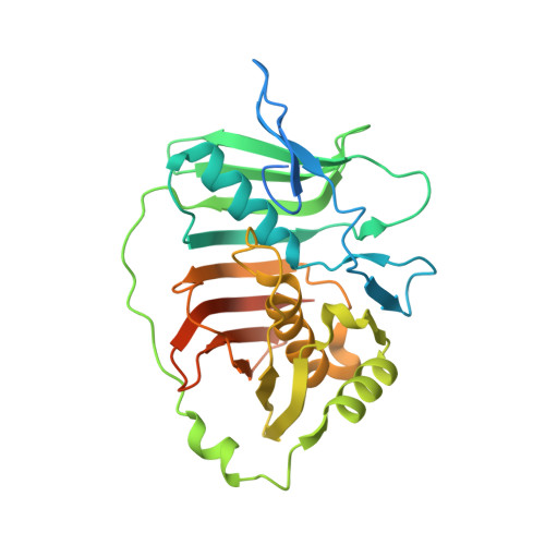 | |
UniProt | |||||
Find proteins for A7Z473 (Bacillus velezensis (strain DSM 23117 / BGSC 10A6 / LMG 26770 / FZB42)) Explore A7Z473 Go to UniProtKB: A7Z473 | |||||
Entity Groups | |||||
| Sequence Clusters | 30% Identity50% Identity70% Identity90% Identity95% Identity100% Identity | ||||
| UniProt Group | A7Z473 | ||||
Sequence AnnotationsExpand | |||||
| |||||
| Length ( Å ) | Angle ( ˚ ) |
|---|---|
| a = 76.05 | α = 90 |
| b = 37.95 | β = 92.28 |
| c = 166.46 | γ = 90 |
| Software Name | Purpose |
|---|---|
| PHENIX | refinement |
| XDS | data reduction |
| XSCALE | data scaling |
| PHASER | phasing |
RCSB PDB (citation) is hosted by
RCSB PDB is a member of the

