5IKB

Crystal structure of the kainate receptor GluK4 ligand binding domain in complex with kainate


Experimental Data Snapshot

  • Method: X-RAY DIFFRACTION
  • Resolution: 2.05 Å
  • R-Value Free: 0.262 
  • R-Value Work: 0.191 
  • R-Value Observed: 0.195 

wwPDB Validation   3D Report Full Report


Ligand Structure Quality Assessment 


This is version 2.1 of the entry. See complete history


Literature

The Structure of a High-Affinity Kainate Receptor: GluK4 Ligand-Binding Domain Crystallized with Kainate.

Kristensen, O.Kristensen, L.B.Mollerud, S.Frydenvang, K.Pickering, D.S.Kastrup, J.S.

(2016) Structure 24: 1582-1589

  • DOI: https://doi.org/10.1016/j.str.2016.06.019
  • Primary Citation of Related Structures:  
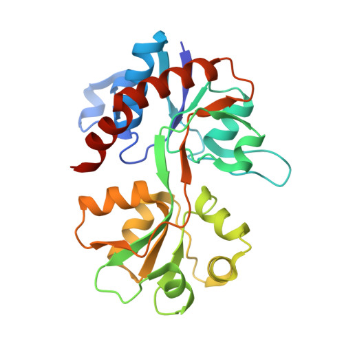
    5IKB

  • PubMed Abstract: 

    Ionotropic glutamate receptors play a key role in fast neurotransmission in the CNS and have been linked to several neurological diseases and disorders. One subfamily is the kainate receptors, which are grouped into low-affinity (GluK1-3) and high-affinity (GluK4-5) receptors based on their affinity for kainate. Although structures of the ligand-binding domain (LBD) of all low-affinity kainate receptors have been reported, no structures of the high-affinity receptor subunits are available. Here, we present the X-ray structure of GluK4-LBD with kainate at 2.05 Å resolution, together with thermofluor and radiolabel binding affinity data. Whereas binding-site residues in GluK4 are most similar to the AMPA receptor subfamily, the domain closure and D1-D2 interlobe contacts induced by kainate are similar to the low-affinity kainate receptor GluK1. These observations provide a likely explanation for the high binding affinity of kainate at GluK4-LBD.


  • Organizational Affiliation

    Department of Drug Design and Pharmacology, Faculty of Health and Medical Sciences, University of Copenhagen, Jagtvej 162, 2100 Copenhagen Ø, Denmark.


Macromolecules
Find similar proteins by:  (by identity cutoff)  |  3D Structure
Entity ID: 1
MoleculeChains Sequence LengthOrganismDetailsImage
Glutamate receptor ionotropic, kainate 4,Glutamate receptor ionotropic, kainate 4257Rattus norvegicusMutation(s): 0 
Gene Names: Grik4
UniProt
Find proteins for Q01812 (Rattus norvegicus)
Explore Q01812 
Go to UniProtKB:  Q01812
Entity Groups  
Sequence Clusters30% Identity50% Identity70% Identity90% Identity95% Identity100% Identity
UniProt GroupQ01812
Sequence Annotations
Expand
  • Reference Sequence
Small Molecules
Binding Affinity Annotations 
IDSourceBinding Affinity
KAI BindingDB:  5IKB Ki: min: 2.29, max: 15 (nM) from 3 assay(s)
Binding MOAD:  5IKB Kd: 1.9 (nM) from 1 assay(s)
Experimental Data & Validation

Experimental Data

  • Method: X-RAY DIFFRACTION
  • Resolution: 2.05 Å
  • R-Value Free: 0.262 
  • R-Value Work: 0.191 
  • R-Value Observed: 0.195 
  • Space Group: C 2 2 21
Unit Cell:
Length ( Å )Angle ( ˚ )
a = 44.5α = 90
b = 69.87β = 90
c = 162.76γ = 90
Software Package:
Software NamePurpose
REFMACrefinement
PHENIXrefinement
xia2data reduction
XDSdata reduction
PHASERphasing
Cootmodel building

Structure Validation

View Full Validation Report


Ligand Structure Quality Assessment 


Entry History & Funding Information

Deposition Data


Funding OrganizationLocationGrant Number
The Lundbeck FoundationDenmark--
E.C. (FP7)Biostruct-X

Revision History  (Full details and data files)

  • Version 1.0: 2016-08-24
    Type: Initial release
  • Version 1.1: 2016-09-14
    Changes: Database references
  • Version 1.2: 2017-08-09
    Changes: Database references
  • Version 2.0: 2019-03-27
    Changes: Atomic model, Data collection, Source and taxonomy
  • Version 2.1: 2024-01-10
    Changes: Data collection, Database references, Refinement description