5IJ7

Structure of Hs/AcPRC2 in complex with a pyridone inhibitor


Experimental Data Snapshot

  • Method: X-RAY DIFFRACTION
  • Resolution: 2.62 Å
  • R-Value Free: 0.238 
  • R-Value Work: 0.191 
  • R-Value Observed: 0.194 

wwPDB Validation   3D Report Full Report


Ligand Structure Quality Assessment 


This is version 1.3 of the entry. See complete history


Literature

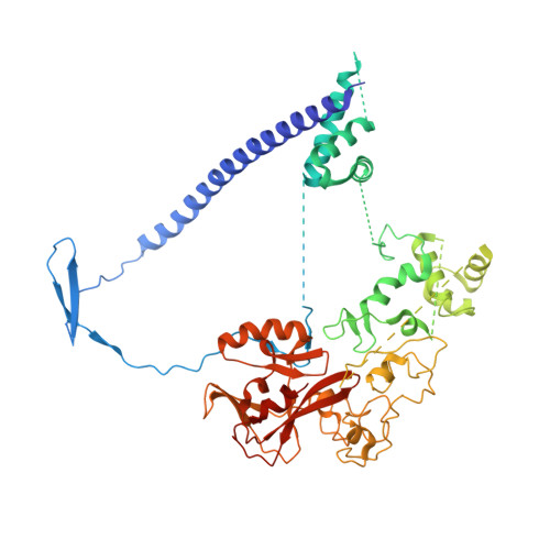
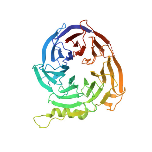
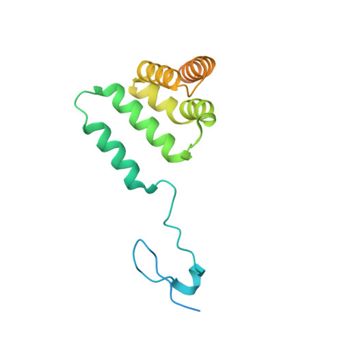
Polycomb repressive complex 2 structure with inhibitor reveals a mechanism of activation and drug resistance.

Brooun, A.Gajiwala, K.S.Deng, Y.L.Liu, W.Bolanos, B.Bingham, P.He, Y.A.Diehl, W.Grable, N.Kung, P.P.Sutton, S.Maegley, K.A.Yu, X.Stewart, A.E.

(2016) Nat Commun 7: 11384-11384

  • DOI: https://doi.org/10.1038/ncomms11384
  • Primary Citation of Related Structures:  
    5IJ7, 5IJ8

  • PubMed Abstract: 

    Polycomb repressive complex 2 (PRC2) mediates gene silencing through chromatin reorganization by methylation of histone H3 lysine 27 (H3K27). Overexpression of the complex and point mutations in the individual subunits of PRC2 have been shown to contribute to tumorigenesis. Several inhibitors of the PRC2 activity have shown efficacy in EZH2-mutated lymphomas and are currently in clinical development, although the molecular basis of inhibitor recognition remains unknown. Here we report the crystal structures of the inhibitor-bound wild-type and Y641N PRC2. The structures illuminate an important role played by a stretch of 17 residues in the N-terminal region of EZH2, we call the activation loop, in the stimulation of the enzyme activity, inhibitor recognition and the potential development of the mutation-mediated drug resistance. The work presented here provides new avenues for the design and development of next-generation PRC2 inhibitors through establishment of a structure-based drug design platform.


  • Organizational Affiliation

    Worldwide Medicinal Chemistry, Worldwide Research and Development, Pfizer Inc., San Diego, California 92121, USA.


Macromolecules
Find similar proteins by:  (by identity cutoff)  |  3D Structure
Entity ID: 1
MoleculeChains Sequence LengthOrganismDetailsImage
Enhancer of Zeste Homolog 2 (EZH2),Histone-lysine N-methyltransferase EZH2
A, B
643Anolis carolinensisHomo sapiens
This entity is chimeric
Mutation(s): 1 
Gene Names: EZH2KMT6
EC: 2.1.1.43
UniProt
Find proteins for G1KPH4 (Anolis carolinensis)
Explore G1KPH4 
Go to UniProtKB:  G1KPH4
Entity Groups  
Sequence Clusters30% Identity50% Identity70% Identity90% Identity95% Identity100% Identity
UniProt GroupG1KPH4
Sequence Annotations
Expand
  • Reference Sequence
Find similar proteins by:  (by identity cutoff)  |  3D Structure
Entity ID: 2
MoleculeChains Sequence LengthOrganismDetailsImage
Polycomb protein EEDC [auth E],
D [auth F]
362Homo sapiensMutation(s): 0 
Gene Names: EED
UniProt & NIH Common Fund Data Resources
Find proteins for O75530 (Homo sapiens)
Explore O75530 
Go to UniProtKB:  O75530
PHAROS:  O75530
GTEx:  ENSG00000074266 
Entity Groups  
Sequence Clusters30% Identity50% Identity70% Identity90% Identity95% Identity100% Identity
UniProt GroupO75530
Sequence Annotations
Expand
  • Reference Sequence
Find similar proteins by:  (by identity cutoff)  |  3D Structure
Entity ID: 3
MoleculeChains Sequence LengthOrganismDetailsImage
Polycomb protein SUZ12E [auth S],
F [auth T]
191Homo sapiensMutation(s): 1 
Gene Names: SUZ12CHET9JJAZ1KIAA0160
UniProt & NIH Common Fund Data Resources
Find proteins for Q15022 (Homo sapiens)
Explore Q15022 
Go to UniProtKB:  Q15022
PHAROS:  Q15022
GTEx:  ENSG00000178691 
Entity Groups  
Sequence Clusters30% Identity50% Identity70% Identity90% Identity95% Identity100% Identity
UniProt GroupQ15022
Sequence Annotations
Expand
  • Reference Sequence
Small Molecules
Ligands 2 Unique
IDChains Name / Formula / InChI Key2D Diagram3D Interactions
6BN
Query on 6BN

Download Ideal Coordinates CCD File 
G [auth A],
O [auth B]
5,8-dichloro-2-[(4-ethyl-6-methyl-2-oxo-1,2-dihydropyridin-3-yl)methyl]-7-({1-[(2R)-2-hydroxypropanoyl]piperidin-4-yl}oxy)-3,4-dihydroisoquinolin-1(2H)-one
C26 H31 Cl2 N3 O5
KBXFBLWYHFRQAH-OAHLLOKOSA-N
ZN
Query on ZN

Download Ideal Coordinates CCD File 
H [auth A]
I [auth A]
J [auth A]
K [auth A]
L [auth A]
H [auth A],
I [auth A],
J [auth A],
K [auth A],
L [auth A],
M [auth A],
N [auth A],
P [auth B],
Q [auth B],
R [auth B],
S [auth B],
T [auth B],
U [auth B],
V [auth B]
ZINC ION
Zn
PTFCDOFLOPIGGS-UHFFFAOYSA-N
Experimental Data & Validation

Experimental Data

  • Method: X-RAY DIFFRACTION
  • Resolution: 2.62 Å
  • R-Value Free: 0.238 
  • R-Value Work: 0.191 
  • R-Value Observed: 0.194 
  • Space Group: P 1 21 1
Unit Cell:
Length ( Å )Angle ( ˚ )
a = 69.575α = 90
b = 115.042β = 102.53
c = 153.033γ = 90
Software Package:
Software NamePurpose
BUSTERrefinement
autoPROCdata scaling
Aimlessdata scaling
autoSHARPphasing

Structure Validation

View Full Validation Report


Ligand Structure Quality Assessment 


Entry History 

Deposition Data

Revision History  (Full details and data files)

  • Version 1.0: 2016-05-04
    Type: Initial release
  • Version 1.1: 2016-05-11
    Changes: Database references
  • Version 1.2: 2017-11-22
    Changes: Derived calculations, Refinement description
  • Version 1.3: 2024-03-06
    Changes: Data collection, Database references, Derived calculations, Refinement description