5IIO

Crystal structure of the DNA polymerase lambda binary complex


Experimental Data Snapshot

  • Method: X-RAY DIFFRACTION
  • Resolution: 2.08 Å
  • R-Value Free: 0.235 
  • R-Value Work: 0.191 
  • R-Value Observed: 0.192 

wwPDB Validation   3D Report Full Report


This is version 1.1 of the entry. See complete history


Literature

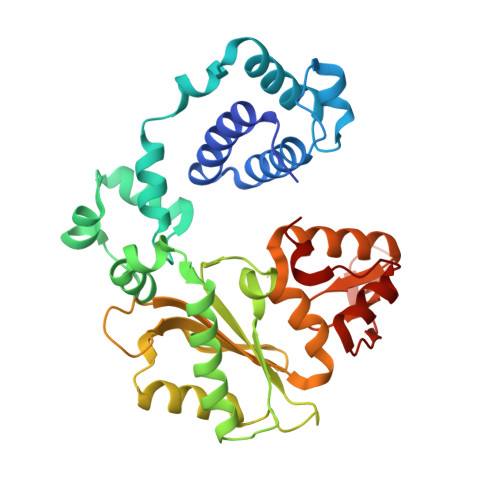
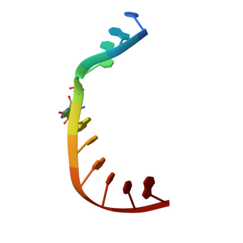
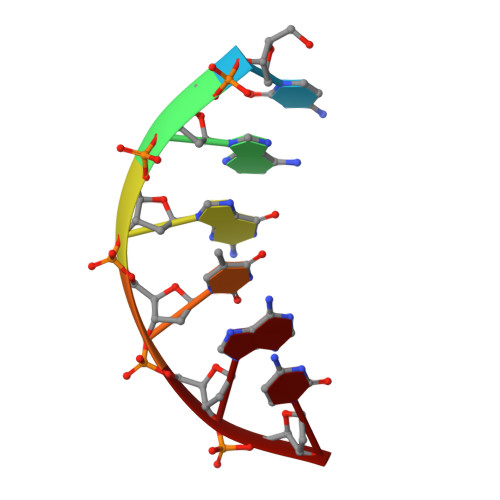
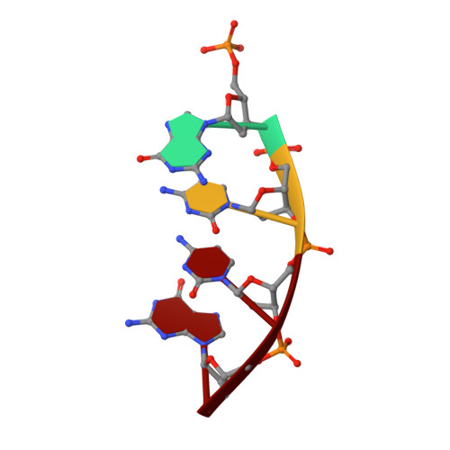
A fidelity mechanism in DNA polymerase lambda promotes error-free bypass of 8-oxo-dG.

Burak, M.J.Guja, K.E.Hambardjieva, E.Derkunt, B.Garcia-Diaz, M.

(2016) EMBO J 35: 2045-2059

  • DOI: https://doi.org/10.15252/embj.201694332
  • Primary Citation of Related Structures:  
    5III, 5IIJ, 5IIK, 5IIL, 5IIM, 5IIN, 5IIO

  • PubMed Abstract: 

    8-oxo-7,8-dihydroxy-2'-deoxyguanosine (8-oxo-dG) has high mutagenic potential as it is prone to mispair with deoxyadenine (dA). In order to maintain genomic integrity, post-replicative 8-oxo-dG:dA mispairs are removed through DNA polymerase lambda (Pol λ)-dependent MUTYH-initiated base excision repair (BER). Here, we describe seven novel crystal structures and kinetic data that fully characterize 8-oxo-dG bypass by Pol λ. We demonstrate that Pol λ has a flexible active site that can tolerate 8-oxo-dG in either the anti- or syn-conformation. Importantly, we show that discrimination against the pro-mutagenic syn-conformation occurs at the extension step and identify the residue responsible for this selectivity. This residue acts as a kinetic switch, shunting repair toward long-patch BER upon correct dCMP incorporation, thus enhancing repair efficiency. Moreover, this switch also provides a potential mechanism to increase repair fidelity of MUTYH-initiated BER.


  • Organizational Affiliation

    Department of Pharmacological Sciences, Stony Brook University, Stony Brook, NY, USA.


Macromolecules

Find similar proteins by:  (by identity cutoff)  |  3D Structure
Entity ID: 4
MoleculeChains Sequence LengthOrganismDetailsImage
DNA polymerase lambdaM [auth A],
N [auth E],
O [auth I],
P [auth M]
334Homo sapiensMutation(s): 0 
Gene Names: POLL
EC: 2.7.7.7 (PDB Primary Data), 4.2.99 (PDB Primary Data)
UniProt & NIH Common Fund Data Resources
Find proteins for Q9UGP5 (Homo sapiens)
Explore Q9UGP5 
Go to UniProtKB:  Q9UGP5
PHAROS:  Q9UGP5
GTEx:  ENSG00000166169 
Entity Groups  
Sequence Clusters30% Identity50% Identity70% Identity90% Identity95% Identity100% Identity
UniProt GroupQ9UGP5
Sequence Annotations
Expand
  • Reference Sequence

Find similar nucleic acids by:  Sequence   |   3D Structure  

Entity ID: 1
MoleculeChains LengthOrganismImage
DNA (5'-D(*CP*GP*GP*CP*(8OG)P*GP*TP*AP*CP*TP*G)-3')A [auth B],
D [auth F],
G [auth J],
J [auth N]
11synthetic construct
Sequence Annotations
Expand
  • Reference Sequence

Find similar nucleic acids by:  Sequence   |   3D Structure  

Entity ID: 2
MoleculeChains LengthOrganismImage
DNA (5'-D(*CP*AP*GP*TP*AP*C)-3')B [auth C],
E [auth G],
H [auth K],
K [auth O]
6synthetic construct
Sequence Annotations
Expand
  • Reference Sequence

Find similar nucleic acids by:  Sequence   |   3D Structure  

Entity ID: 3
MoleculeChains LengthOrganismImage
DNA (5'-D(P*GP*CP*CP*G)-3')C [auth D],
F [auth H],
I [auth L],
L [auth P]
4synthetic construct
Sequence Annotations
Expand
  • Reference Sequence
Experimental Data & Validation

Experimental Data

  • Method: X-RAY DIFFRACTION
  • Resolution: 2.08 Å
  • R-Value Free: 0.235 
  • R-Value Work: 0.191 
  • R-Value Observed: 0.192 
  • Space Group: P 21 21 2
Unit Cell:
Length ( Å )Angle ( ˚ )
a = 191.423α = 90
b = 98.98β = 90
c = 105.034γ = 90
Software Package:
Software NamePurpose
PHENIXrefinement
XDSdata reduction
Aimlessdata scaling
PHASERphasing

Structure Validation

View Full Validation Report


Entry History 

Deposition Data

Revision History  (Full details and data files)

  • Version 1.0: 2016-08-17
    Type: Initial release
  • Version 1.1: 2016-09-28
    Changes: Database references