


Crystal structure of mouse CARM1 in complex with SAH at 1.8 Angstroms resolution
Cura, V., Marechal, N., Mailliot, J., Troffer-Charlier, N., Wurtz, J.M., Bonnefond, L., Cavarelli, J.To be published.
Experimental Data Snapshot
Entity ID: 1 | |||||
|---|---|---|---|---|---|
| Molecule | Chains | Sequence Length | Organism | Details | Image |
| Histone-arginine methyltransferase CARM1 | 361 | Mus musculus | Mutation(s): 0 Gene Names: Carm1, Prmt4 EC: 2.1.1.319 | 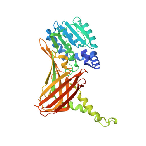 | |
UniProt & NIH Common Fund Data Resources | |||||
Find proteins for Q9WVG6 (Mus musculus) Explore Q9WVG6 Go to UniProtKB: Q9WVG6 | |||||
IMPC: MGI:1913208 | |||||
Entity Groups | |||||
| Sequence Clusters | 30% Identity50% Identity70% Identity90% Identity95% Identity100% Identity | ||||
| UniProt Group | Q9WVG6 | ||||
Sequence AnnotationsExpand | |||||
| |||||
| Ligands 6 Unique | |||||
|---|---|---|---|---|---|
| ID | Chains | Name / Formula / InChI Key | 2D Diagram | 3D Interactions | |
| SAH Query on SAH | BA [auth D], E [auth A], L [auth B], T [auth C] | S-ADENOSYL-L-HOMOCYSTEINE C14 H20 N6 O5 S ZJUKTBDSGOFHSH-WFMPWKQPSA-N |  | ||
| PE8 Query on PE8 | Q [auth B] | 3,6,9,12,15,18,21-HEPTAOXATRICOSANE-1,23-DIOL C16 H34 O9 GLZWNFNQMJAZGY-UHFFFAOYSA-N |  | ||
| PGE Query on PGE | K [auth A] | TRIETHYLENE GLYCOL C6 H14 O4 ZIBGPFATKBEMQZ-UHFFFAOYSA-N |  | ||
| PEG Query on PEG | H [auth A] I [auth A] J [auth A] P [auth B] X [auth C] | DI(HYDROXYETHYL)ETHER C4 H10 O3 MTHSVFCYNBDYFN-UHFFFAOYSA-N |  | ||
| DXE Query on DXE | AA [auth C], R [auth B], S [auth B], Z [auth C] | 1,2-DIMETHOXYETHANE C4 H10 O2 XTHFKEDIFFGKHM-UHFFFAOYSA-N |  | ||
| EDO Query on EDO | CA [auth D] DA [auth D] EA [auth D] F [auth A] G [auth A] | 1,2-ETHANEDIOL C2 H6 O2 LYCAIKOWRPUZTN-UHFFFAOYSA-N |  | ||
| Length ( Å ) | Angle ( ˚ ) |
|---|---|
| a = 74.628 | α = 90 |
| b = 97.92 | β = 90 |
| c = 204.955 | γ = 90 |
| Software Name | Purpose |
|---|---|
| PHENIX | refinement |
| XDS | data reduction |
| Aimless | data scaling |
| Funding Organization | Location | Grant Number |
|---|---|---|
| France | -- |
RCSB PDB (citation) is hosted by
RCSB PDB is a member of the

