


Crystal structure of a short chain dehydrogenase/reductase SDR from Burkholderia phymatum in complex with NAD
Mayclin, S.J., Dranow, D.M., Lorimer, D.D., Edwards, T.E.To be published.
Experimental Data Snapshot
Entity ID: 1 | |||||
|---|---|---|---|---|---|
| Molecule | Chains | Sequence Length | Organism | Details | Image |
| Short-chain dehydrogenase/reductase SDR | 297 | Paraburkholderia phymatum STM815 | Mutation(s): 0 Gene Names: Bphy_1038 | 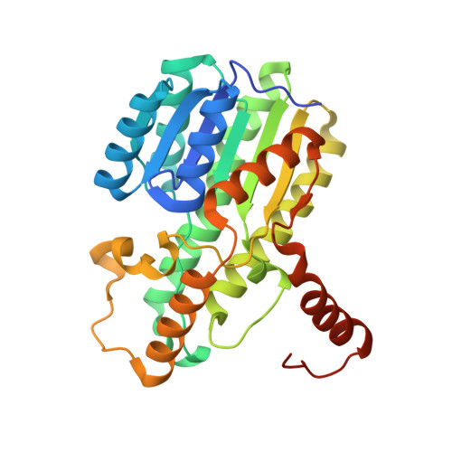 | |
UniProt | |||||
Find proteins for B2JGP2 (Paraburkholderia phymatum (strain DSM 17167 / CIP 108236 / LMG 21445 / STM815)) Explore B2JGP2 Go to UniProtKB: B2JGP2 | |||||
Entity Groups | |||||
| Sequence Clusters | 30% Identity50% Identity70% Identity90% Identity95% Identity100% Identity | ||||
| UniProt Group | B2JGP2 | ||||
Sequence AnnotationsExpand | |||||
| |||||
| Ligands 2 Unique | |||||
|---|---|---|---|---|---|
| ID | Chains | Name / Formula / InChI Key | 2D Diagram | 3D Interactions | |
| NAD Query on NAD | D [auth A], F [auth B], H [auth C] | NICOTINAMIDE-ADENINE-DINUCLEOTIDE C21 H27 N7 O14 P2 BAWFJGJZGIEFAR-NNYOXOHSSA-N |  | ||
| ACT Query on ACT | E [auth A], G [auth B], I [auth C] | ACETATE ION C2 H3 O2 QTBSBXVTEAMEQO-UHFFFAOYSA-M |  | ||
| Length ( Å ) | Angle ( ˚ ) |
|---|---|
| a = 83.52 | α = 90 |
| b = 187.79 | β = 90 |
| c = 108.26 | γ = 90 |
| Software Name | Purpose |
|---|---|
| PHENIX | refinement |
| XSCALE | data scaling |
| MOLREP | phasing |
| PDB_EXTRACT | data extraction |
| Coot | model building |
| XDS | data reduction |
RCSB PDB (citation) is hosted by
RCSB PDB is a member of the

