


Crystal Structure of a Cysteine Synthase from Brucella suis
Dranow, D.M., Lorimer, D., Edwards, T.E.To be published.
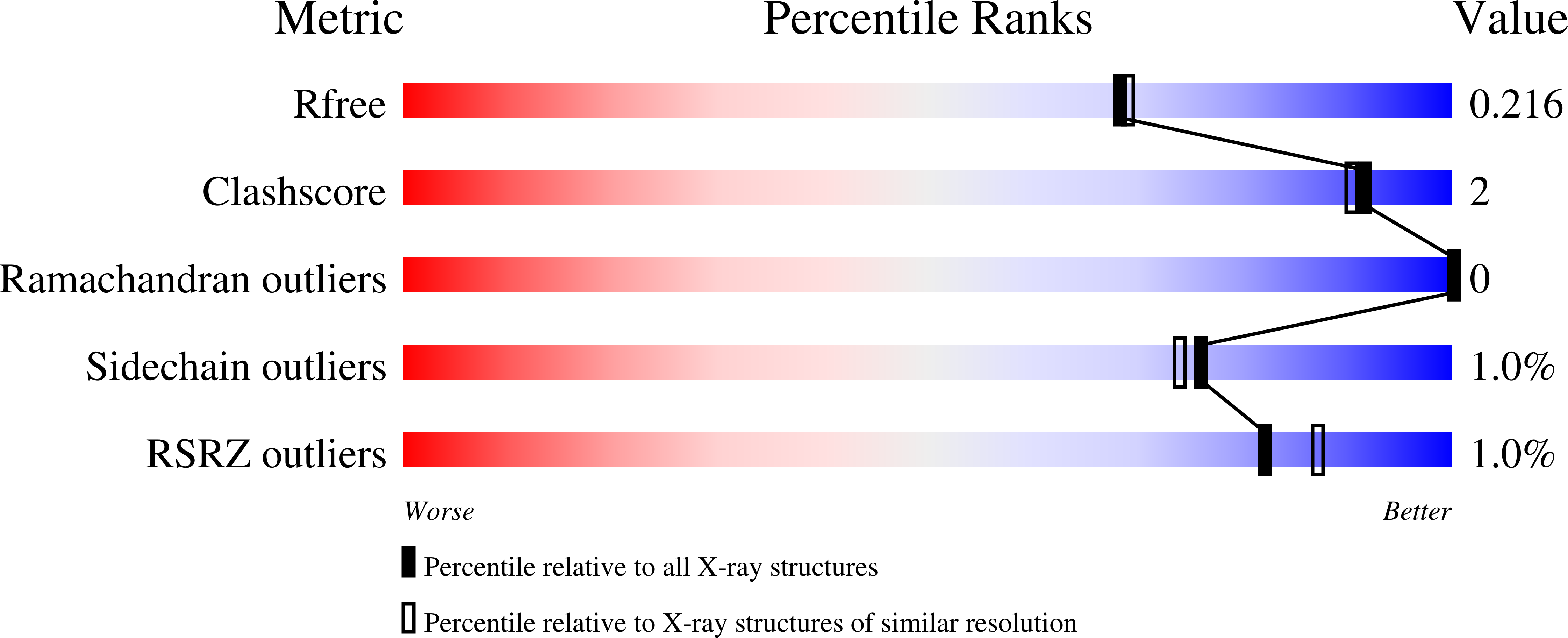
Experimental Data Snapshot
wwPDB Validation 3D Report Full Report
Entity ID: 1 | |||||
|---|---|---|---|---|---|
| Molecule | Chains | Sequence Length | Organism | Details | Image |
| Cysteine synthase A | 363 | Brucella suis 1330 | Mutation(s): 0 Gene Names: BS1330_I1049 EC: 2.5.1.47 |  | |
UniProt | |||||
Find proteins for A0A0H3G350 (Brucella suis biovar 1 (strain 1330)) Explore A0A0H3G350 Go to UniProtKB: A0A0H3G350 | |||||
Entity Groups | |||||
| Sequence Clusters | 30% Identity50% Identity70% Identity90% Identity95% Identity100% Identity | ||||
| UniProt Group | A0A0H3G350 | ||||
Sequence AnnotationsExpand | |||||
| |||||
| Ligands 1 Unique | |||||
|---|---|---|---|---|---|
| ID | Chains | Name / Formula / InChI Key | 2D Diagram | 3D Interactions | |
| MLI Query on MLI | C [auth A], D [auth A], E [auth B] | MALONATE ION C3 H2 O4 OFOBLEOULBTSOW-UHFFFAOYSA-L |  | ||
| Modified Residues 1 Unique | |||||
|---|---|---|---|---|---|
| ID | Chains | Type | Formula | 2D Diagram | Parent |
| LLP Query on LLP | A, B | L-PEPTIDE LINKING | C14 H22 N3 O7 P |  | LYS |
| Length ( Å ) | Angle ( ˚ ) |
|---|---|
| a = 60.02 | α = 90 |
| b = 71.86 | β = 90 |
| c = 201.6 | γ = 90 |
| Software Name | Purpose |
|---|---|
| XSCALE | data scaling |
| PHENIX | refinement |
| PDB_EXTRACT | data extraction |
| XDS | data reduction |
RCSB PDB (citation) is hosted by
RCSB PDB is a member of the

