


Crystal Structure of plasmepsin IV
Asojo, O.A.To be published.
Experimental Data Snapshot
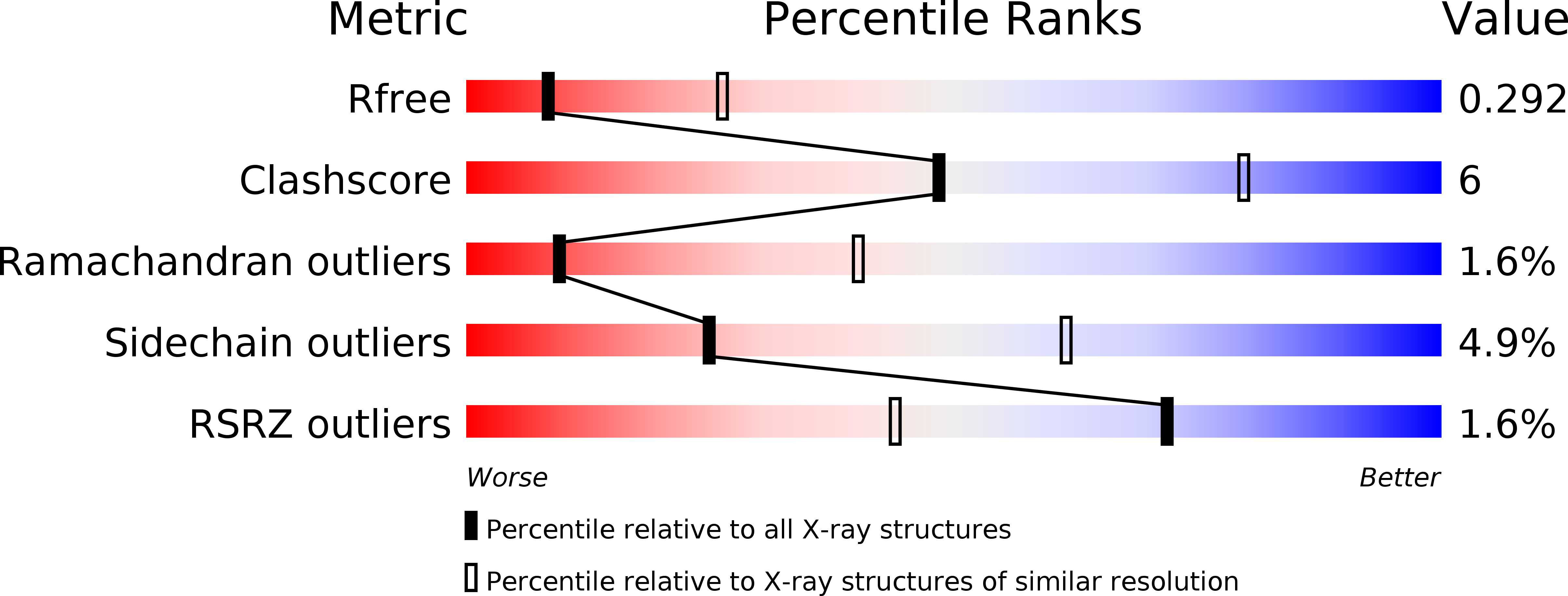
wwPDB Validation 3D Report Full Report
Entity ID: 1 | |||||
|---|---|---|---|---|---|
| Molecule | Chains | Sequence Length | Organism | Details | Image |
| Plasmepsin IV | A [auth B], B [auth A] | 328 | Plasmodium falciparum 7G8 | Mutation(s): 0 Gene Names: PFBG_05102 | 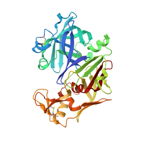 |
UniProt | |||||
Find proteins for W7FF86 (Plasmodium falciparum (isolate 7G8)) Explore W7FF86 Go to UniProtKB: W7FF86 | |||||
Entity Groups | |||||
| Sequence Clusters | 30% Identity50% Identity70% Identity90% Identity95% Identity100% Identity | ||||
| UniProt Group | W7FF86 | ||||
Sequence AnnotationsExpand | |||||
| |||||
Find similar proteins by: Sequence | 3D Structure
Entity ID: 2 | |||||
|---|---|---|---|---|---|
| Molecule | Chains | Sequence Length | Organism | Details | Image |
| Pepstatin A | 6 | Actinomyces | Mutation(s): 0 | 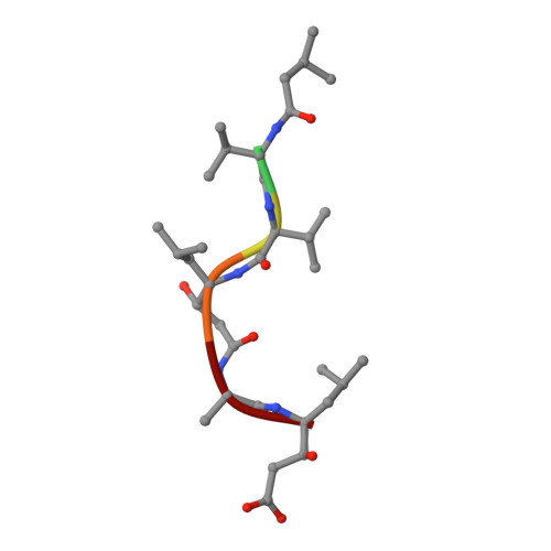 | |
Sequence AnnotationsExpand | |||||
| |||||
Entity ID: 2 | |||||
|---|---|---|---|---|---|
| ID | Chains | Name | Type/Class | 2D Diagram | 3D Interactions |
| PRD_000557 Query on PRD_000557 | C, D | Pepstatin | Oligopeptide / Enzyme inhibitor |  | |
| Length ( Å ) | Angle ( ˚ ) |
|---|---|
| a = 103.42 | α = 90 |
| b = 103.42 | β = 90 |
| c = 123.69 | γ = 120 |
| Software Name | Purpose |
|---|---|
| REFMAC | refinement |
| iMOSFLM | data reduction |
| SCALA | data scaling |
| PHASER | phasing |
RCSB PDB (citation) is hosted by
RCSB PDB is a member of the

