5I28

Azurin T30R1, crystal form II


Experimental Data Snapshot

  • Method: X-RAY DIFFRACTION
  • Resolution: 1.95 Å
  • R-Value Free: 0.271 
  • R-Value Work: 0.212 
  • R-Value Observed: 0.212 

wwPDB Validation   3D Report Full Report


This is version 1.2 of the entry. See complete history


Literature

Determination of nitroxide spin label conformations via PELDOR and X-ray crystallography.

Abdullin, D.Hagelueken, G.Schiemann, O.

(2016) Phys Chem Chem Phys 18: 10428-10437

  • DOI: https://doi.org/10.1039/c6cp01307d
  • Primary Citation of Related Structures:  
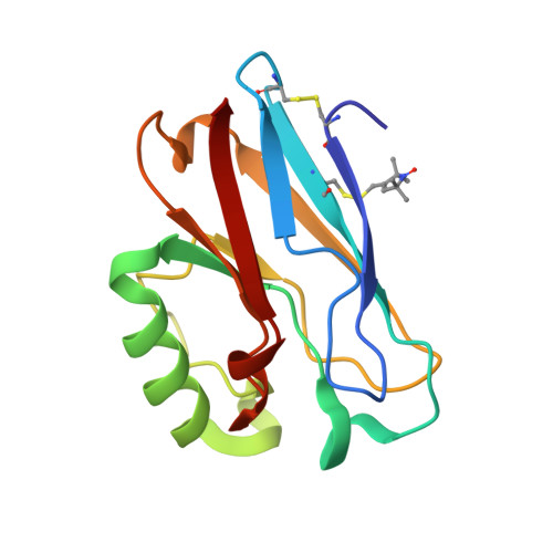
    5I26, 5I28

  • PubMed Abstract: 

    Pulsed electron-electron double resonance (PELDOR or DEER) in combination with site-directed spin labelling has emerged as an important method for measuring nanometer distance constraints that are used to obtain coarse-grained structures of biomolecules or to follow their conformational changes. Translating measured spin-spin distances between spin labels into structural information requires taking the conformational flexibility of spin label side chains into account. Here, we present an analysis of orientation selective PELDOR data recorded on six singly MTSSL-labelled azurin mutants. The analysis yielded conformational MTSSL ensembles, which are considerably narrower than those predicted using in silico spin labeling methods but match well with spin label conformations found in the corresponding crystal structures. The possible reasons and consequences for predicting spin label conformers in the fold of biomolecules are discussed.


  • Organizational Affiliation

    Institute of Physical and Theoretical Chemistry, University of Bonn, Wegelerstr. 12, 53115 Bonn, Germany. schiemann@pc.uni-bonn.de.


Macromolecules
Find similar proteins by:  (by identity cutoff)  |  3D Structure
Entity ID: 1
MoleculeChains Sequence LengthOrganismDetailsImage
Azurin
A, B, C, D, E
A, B, C, D, E, F, G, H, I, J, K, L, M, N, O, P
128Pseudomonas aeruginosaMutation(s): 1 
Gene Names: azuPA4922
UniProt
Find proteins for P00282 (Pseudomonas aeruginosa (strain ATCC 15692 / DSM 22644 / CIP 104116 / JCM 14847 / LMG 12228 / 1C / PRS 101 / PAO1))
Explore P00282 
Go to UniProtKB:  P00282
Entity Groups  
Sequence Clusters30% Identity50% Identity70% Identity90% Identity95% Identity100% Identity
UniProt GroupP00282
Sequence Annotations
Expand
  • Reference Sequence
Small Molecules
Ligands 2 Unique
IDChains Name / Formula / InChI Key2D Diagram3D Interactions
GOL
Query on GOL

Download Ideal Coordinates CCD File 
R [auth A]GLYCEROL
C3 H8 O3
PEDCQBHIVMGVHV-UHFFFAOYSA-N
CU
Query on CU

Download Ideal Coordinates CCD File 
AA [auth J]
BA [auth K]
CA [auth L]
DA [auth M]
EA [auth N]
AA [auth J],
BA [auth K],
CA [auth L],
DA [auth M],
EA [auth N],
FA [auth O],
GA [auth P],
Q [auth A],
S [auth B],
T [auth C],
U [auth D],
V [auth E],
W [auth F],
X [auth G],
Y [auth H],
Z [auth I]
COPPER (II) ION
Cu
JPVYNHNXODAKFH-UHFFFAOYSA-N
Modified Residues  1 Unique
IDChains TypeFormula2D DiagramParent
R1A
Query on R1A
A, B, C, D, E
A, B, C, D, E, F, G, H, I, J, K, L, M, N, O, P
L-PEPTIDE LINKINGC12 H21 N2 O3 S2CYS
Experimental Data & Validation

Experimental Data

  • Method: X-RAY DIFFRACTION
  • Resolution: 1.95 Å
  • R-Value Free: 0.271 
  • R-Value Work: 0.212 
  • R-Value Observed: 0.212 
  • Space Group: C 1 2 1
Unit Cell:
Length ( Å )Angle ( ˚ )
a = 242.478α = 90
b = 105.56β = 114.42
c = 100.051γ = 90
Software Package:
Software NamePurpose
PHENIXrefinement
XDSdata reduction
XDSdata scaling
PHASERphasing

Structure Validation

View Full Validation Report


Entry History 

Deposition Data

Revision History  (Full details and data files)

  • Version 1.0: 2016-04-13
    Type: Initial release
  • Version 1.1: 2016-04-20
    Changes: Database references
  • Version 1.2: 2024-01-10
    Changes: Data collection, Database references, Derived calculations, Refinement description