Arylfluorosulfates Inactivate Intracellular Lipid Binding Protein(s) through Chemoselective SuFEx Reaction with a Binding Site Tyr Residue.
Chen, W., Dong, J., Plate, L., Mortenson, D.E., Brighty, G.J., Li, S., Liu, Y., Galmozzi, A., Lee, P.S., Hulce, J.J., Cravatt, B.F., Saez, E., Powers, E.T., Wilson, I.A., Sharpless, K.B., Kelly, J.W.(2016) J Am Chem Soc 138: 7353-7364
- PubMed: 27191344
- DOI: https://doi.org/10.1021/jacs.6b02960
- Primary Citation of Related Structures:
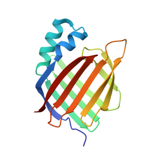
5HZQ - PubMed Abstract:
Arylfluorosulfates have appeared only rarely in the literature and have not been explored as probes for covalent conjugation to proteins, possibly because they were assumed to possess high reactivity, as with other sulfur(VI) halides. However, we find that arylfluorosulfates become reactive only under certain circumstances, e.g., when fluoride displacement by a nucleophile is facilitated. Herein, we explore the reactivity of structurally simple arylfluorosulfates toward the proteome of human cells. We demonstrate that the protein reactivity of arylfluorosulfates is lower than that of the corresponding aryl sulfonyl fluorides, which are better characterized with regard to proteome reactivity. We discovered that simple hydrophobic arylfluorosulfates selectively react with a few members of the intracellular lipid binding protein (iLBP) family. A central function of iLBPs is to deliver small-molecule ligands to nuclear hormone receptors. Arylfluorosulfate probe 1 reacts with a conserved tyrosine residue in the ligand-binding site of a subset of iLBPs. Arylfluorosulfate probes 3 and 4, featuring a biphenyl core, very selectively and efficiently modify cellular retinoic acid binding protein 2 (CRABP2), both in vitro and in living cells. The X-ray crystal structure of the CRABP2-4 conjugate, when considered together with binding site mutagenesis experiments, provides insight into how CRABP2 might activate arylfluorosulfates toward site-specific reaction. Treatment of breast cancer cells with probe 4 attenuates nuclear hormone receptor activity mediated by retinoic acid, an endogenous client lipid of CRABP2. Our findings demonstrate that arylfluorosulfates can selectively target single iLBPs, making them useful for understanding iLBP function.
Organizational Affiliation:
Department of Chemistry and §Department of Molecular and Experimental Medicine, ∥Department of Chemical Physiology, ⊥Department of Integrative Structural and Computational Biology, #The Skaggs Institute for Chemical Biology, The Scripps Research Institute , La Jolla, California 92037, United States.