5HYK

Crystal structure of the complex PPARalpha/AL26-29


Experimental Data Snapshot

  • Method: X-RAY DIFFRACTION
  • Resolution: 1.83 Å
  • R-Value Free: 0.262 
  • R-Value Work: 0.211 
  • R-Value Observed: 0.214 

wwPDB Validation   3D Report Full Report


Ligand Structure Quality Assessment 


This is version 2.0 of the entry. See complete history


Literature

Structural basis for PPAR partial or full activation revealed by a novel ligand binding mode.

Capelli, D.Cerchia, C.Montanari, R.Loiodice, F.Tortorella, P.Laghezza, A.Cervoni, L.Pochetti, G.Lavecchia, A.

(2016) Sci Rep 6: 34792-34792

  • DOI: https://doi.org/10.1038/srep34792
  • Primary Citation of Related Structures:  
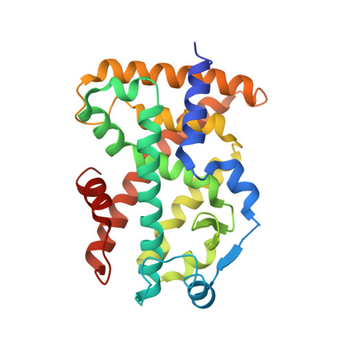
    5HYK, 5HZC

  • PubMed Abstract: 

    The peroxisome proliferator-activated receptors (PPARs) are nuclear receptors involved in the regulation of the metabolic homeostasis and therefore represent valuable therapeutic targets for the treatment of metabolic diseases. The development of more balanced drugs interacting with PPARs, devoid of the side-effects showed by the currently marketed PPARγ full agonists, is considered the major challenge for the pharmaceutical companies. Here we present a structure-based virtual screening approach that let us identify a novel PPAR pan-agonist with a very attractive activity profile and its crystal structure in the complex with PPARα and PPARγ, respectively. In PPARα this ligand occupies a new pocket whose filling is allowed by the ligand-induced switching of the F273 side chain from a closed to an open conformation. The comparison between this pocket and the corresponding cavity in PPARγ provides a rationale for the different activation of the ligand towards PPARα and PPARγ, suggesting a novel basis for ligand design.


  • Organizational Affiliation

    Istituto di Cristallografia, Consiglio Nazionale delle Ricerche, Via Salaria Km. 29, 300, 00015 Monterotondo Stazione, Roma, Italy.


Macromolecules
Find similar proteins by:  (by identity cutoff)  |  3D Structure
Entity ID: 1
MoleculeChains Sequence LengthOrganismDetailsImage
Peroxisome proliferator-activated receptor alpha272Homo sapiensMutation(s): 0 
Gene Names: PPARANR1C1PPAR
UniProt & NIH Common Fund Data Resources
Find proteins for Q07869 (Homo sapiens)
Explore Q07869 
Go to UniProtKB:  Q07869
PHAROS:  Q07869
GTEx:  ENSG00000186951 
Entity Groups  
Sequence Clusters30% Identity50% Identity70% Identity90% Identity95% Identity100% Identity
UniProt GroupQ07869
Sequence Annotations
Expand
  • Reference Sequence
Small Molecules
Ligands 1 Unique
IDChains Name / Formula / InChI Key2D Diagram3D Interactions
65W
Query on 65W

Download Ideal Coordinates CCD File 
B [auth A]2-methyl-2-[4-(naphthalen-1-yl)phenoxy]propanoic acid
C20 H18 O3
JTWKEZBNKHBDAW-UHFFFAOYSA-N
Experimental Data & Validation

Experimental Data

  • Method: X-RAY DIFFRACTION
  • Resolution: 1.83 Å
  • R-Value Free: 0.262 
  • R-Value Work: 0.211 
  • R-Value Observed: 0.214 
  • Space Group: P 41 21 2
Unit Cell:
Length ( Å )Angle ( ˚ )
a = 63.65α = 90
b = 63.65β = 90
c = 126γ = 90
Software Package:
Software NamePurpose
PHENIXrefinement
iMOSFLMdata reduction
SCALAdata scaling
AMoREphasing

Structure Validation

View Full Validation Report


Ligand Structure Quality Assessment 


Entry History 

Deposition Data

Revision History  (Full details and data files)

  • Version 1.0: 2016-11-23
    Type: Initial release
  • Version 2.0: 2024-01-10
    Changes: Atomic model, Data collection, Database references, Refinement description