5HXY

Crystal structure of XerA recombinase


Experimental Data Snapshot

  • Method: X-RAY DIFFRACTION
  • Resolution: 2.50 Å
  • R-Value Free: 0.243 
  • R-Value Work: 0.188 
  • R-Value Observed: 0.191 

wwPDB Validation   3D Report Full Report


This is version 1.1 of the entry. See complete history


Literature

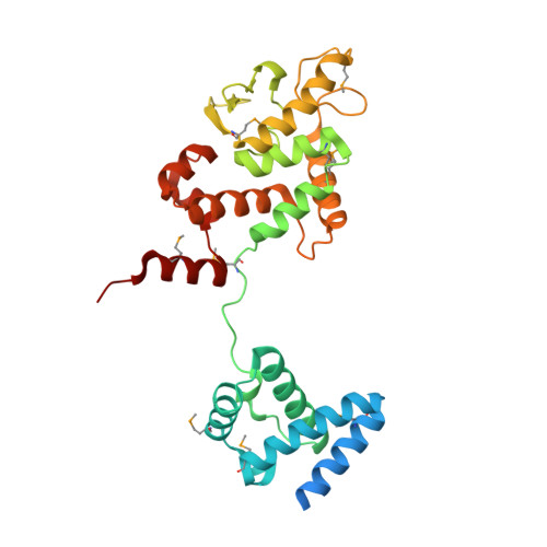
Crystal structure of Thermoplasma acidophilum XerA recombinase shows large C-shape clamp conformation and cis-cleavage mode for nucleophilic tyrosine

Jo, C.H.Kim, J.Han, A.R.Park, S.Y.Hwang, K.Y.Nam, K.H.

(2016) FEBS Lett 590: 848-856

  • DOI: https://doi.org/10.1002/1873-3468.12109
  • Primary Citation of Related Structures:  
    5HXY

  • PubMed Abstract: 

    Site-specific Xer recombination plays a pivotal role in reshuffling genetic information. Here, we report the 2.5 Å crystal structure of XerA from the archaean Thermoplasma acidophilum. Crystallographic data reveal a uniquely open conformational state, resulting in a C-shaped clamp with an angle of ~ 48° and a distance of 57 Å between the core-binding and the catalytic domains. The catalytic nucleophile, Tyr264, is positioned in cis-cleavage mode by XerA's C-term tail that interacts with the CAT domain of a neighboring monomer without DNA substrate. Structural comparisons of tyrosine recombinases elucidate the dynamics of Xer recombinase.


  • Organizational Affiliation

    Division of Biotechnology, College of Life Sciences and Biotechnology, Korea University, Seoul, Korea.


Macromolecules
Find similar proteins by:  (by identity cutoff)  |  3D Structure
Entity ID: 1
MoleculeChains Sequence LengthOrganismDetailsImage
Tyrosine recombinase XerA
A, B, C, D, E
A, B, C, D, E, F
317Thermoplasma acidophilum DSM 1728Mutation(s): 0 
Gene Names: xerATa1314
UniProt
Find proteins for Q9HIM5 (Thermoplasma acidophilum (strain ATCC 25905 / DSM 1728 / JCM 9062 / NBRC 15155 / AMRC-C165))
Explore Q9HIM5 
Go to UniProtKB:  Q9HIM5
Entity Groups  
Sequence Clusters30% Identity50% Identity70% Identity90% Identity95% Identity100% Identity
UniProt GroupQ9HIM5
Sequence Annotations
Expand
  • Reference Sequence
Small Molecules
Ligands 1 Unique
IDChains Name / Formula / InChI Key2D Diagram3D Interactions
PO4
Query on PO4

Download Ideal Coordinates CCD File 
G [auth A]
H [auth A]
I [auth A]
J [auth A]
K [auth B]
G [auth A],
H [auth A],
I [auth A],
J [auth A],
K [auth B],
L [auth B],
M [auth C],
N [auth C],
O [auth C],
P [auth D],
Q [auth D],
R [auth E],
S [auth E],
T [auth F],
U [auth F]
PHOSPHATE ION
O4 P
NBIIXXVUZAFLBC-UHFFFAOYSA-K
Modified Residues  1 Unique
IDChains TypeFormula2D DiagramParent
MSE
Query on MSE
A, B, C, D, E
A, B, C, D, E, F
L-PEPTIDE LINKINGC5 H11 N O2 SeMET
Experimental Data & Validation

Experimental Data

  • Method: X-RAY DIFFRACTION
  • Resolution: 2.50 Å
  • R-Value Free: 0.243 
  • R-Value Work: 0.188 
  • R-Value Observed: 0.191 
  • Space Group: C 1 2 1
Unit Cell:
Length ( Å )Angle ( ˚ )
a = 182.86α = 90
b = 105.968β = 110.01
c = 115.586γ = 90
Software Package:
Software NamePurpose
REFMACrefinement
HKL-2000data collection
HKL-2000data scaling
SOLVEphasing
RESOLVEmodel building
PDB_EXTRACTdata extraction

Structure Validation

View Full Validation Report


Entry History & Funding Information

Deposition Data


Funding OrganizationLocationGrant Number
National Research Foundation of Korea (NRF) grant funded by the Korea government (MEST)Korea, Republic OfNRF-2014R1A1A2059440
National Research Foundation of Korea (NRF) grant funded by the Korea government (MEST)Korea, Republic OfNRF-2014R1212513

Revision History  (Full details and data files)

  • Version 1.0: 2017-02-01
    Type: Initial release
  • Version 1.1: 2020-02-19
    Changes: Data collection