5HVK

Crystal structure of LIMK1 mutant D460N in complex with full-length cofilin-1


Experimental Data Snapshot

  • Method: X-RAY DIFFRACTION
  • Resolution: 3.50 Å
  • R-Value Free: 0.310 
  • R-Value Work: 0.273 
  • R-Value Observed: 0.275 

wwPDB Validation   3D Report Full Report


Ligand Structure Quality Assessment 


This is version 1.4 of the entry. See complete history


Literature

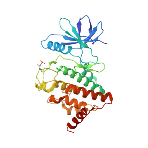
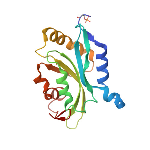
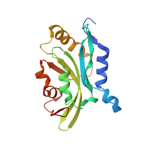
Structural Basis for Noncanonical Substrate Recognition of Cofilin/ADF Proteins by LIM Kinases.

Hamill, S.Lou, H.J.Turk, B.E.Boggon, T.J.

(2016) Mol Cell 62: 397-408

  • DOI: https://doi.org/10.1016/j.molcel.2016.04.001
  • Primary Citation of Related Structures:  
    5HVJ, 5HVK

  • PubMed Abstract: 

    Cofilin/actin-depolymerizing factor (ADF) proteins are critical nodes that relay signals from protein kinase cascades to the actin cytoskeleton, in particular through site-specific phosphorylation at residue Ser3. This is important for regulation of the roles of cofilin in severing and stabilizing actin filaments. Consequently, cofilin/ADF Ser3 phosphorylation is tightly controlled as an almost exclusive substrate for LIM kinases. Here we determine the LIMK1:cofilin-1 co-crystal structure. We find an interface that is distinct from canonical kinase-substrate interactions. We validate this previously unobserved mechanism for high-fidelity kinase-substrate recognition by in vitro kinase assays, examination of cofilin phosphorylation in mammalian cells, and functional analysis in S. cerevisiae. The interface is conserved across all LIM kinases. Remarkably, we also observe both pre- and postphosphotransfer states in the same crystal lattice. This study therefore provides a molecular understanding of how kinase-substrate recognition acts as a gatekeeper to regulate actin cytoskeletal dynamics.


  • Organizational Affiliation

    Department of Pharmacology, Yale University School of Medicine, New Haven, CT 06520, USA.


Macromolecules
Find similar proteins by:  (by identity cutoff)  |  3D Structure
Entity ID: 1
MoleculeChains Sequence LengthOrganismDetailsImage
LIM domain kinase 1
A, C
315Homo sapiensMutation(s): 1 
Gene Names: LIMK1LIMK
EC: 2.7.11.1
UniProt & NIH Common Fund Data Resources
Find proteins for P53667 (Homo sapiens)
Explore P53667 
Go to UniProtKB:  P53667
PHAROS:  P53667
GTEx:  ENSG00000106683 
Entity Groups  
Sequence Clusters30% Identity50% Identity70% Identity90% Identity95% Identity100% Identity
UniProt GroupP53667
Sequence Annotations
Expand
  • Reference Sequence
Find similar proteins by:  (by identity cutoff)  |  3D Structure
Entity ID: 2
MoleculeChains Sequence LengthOrganismDetailsImage
Cofilin-1165Homo sapiensMutation(s): 1 
Gene Names: CFL1CFL
UniProt & NIH Common Fund Data Resources
Find proteins for P23528 (Homo sapiens)
Explore P23528 
Go to UniProtKB:  P23528
PHAROS:  P23528
GTEx:  ENSG00000172757 
Entity Groups  
Sequence Clusters30% Identity50% Identity70% Identity90% Identity95% Identity100% Identity
UniProt GroupP23528
Sequence Annotations
Expand
  • Reference Sequence
Find similar proteins by:  (by identity cutoff)  |  3D Structure
Entity ID: 3
MoleculeChains Sequence LengthOrganismDetailsImage
Cofilin-1165Homo sapiensMutation(s): 1 
Gene Names: CFL1CFL
UniProt & NIH Common Fund Data Resources
Find proteins for P23528 (Homo sapiens)
Explore P23528 
Go to UniProtKB:  P23528
PHAROS:  P23528
GTEx:  ENSG00000172757 
Entity Groups  
Sequence Clusters30% Identity50% Identity70% Identity90% Identity95% Identity100% Identity
UniProt GroupP23528
Sequence Annotations
Expand
  • Reference Sequence
Small Molecules
Modified Residues  2 Unique
IDChains TypeFormula2D DiagramParent
TPO
Query on TPO
A, C
L-PEPTIDE LINKINGC4 H10 N O6 PTHR
SEP
Query on SEP
B
L-PEPTIDE LINKINGC3 H8 N O6 PSER
Experimental Data & Validation

Experimental Data

  • Method: X-RAY DIFFRACTION
  • Resolution: 3.50 Å
  • R-Value Free: 0.310 
  • R-Value Work: 0.273 
  • R-Value Observed: 0.275 
  • Space Group: P 21 21 21
Unit Cell:
Length ( Å )Angle ( ˚ )
a = 81.116α = 90
b = 102.283β = 90
c = 141.9γ = 90
Software Package:
Software NamePurpose
PHENIXrefinement
HKL-2000data reduction
HKL-2000data scaling
Cootmodel building
PHENIXphasing

Structure Validation

View Full Validation Report


Ligand Structure Quality Assessment 


Entry History & Funding Information

Deposition Data


Funding OrganizationLocationGrant Number
National Institutes of Health/National Institute of General Medical Sciences (NIH/NIGMS)United StatesR01GM102262
National Institutes of Health/National Institute of General Medical Sciences (NIH/NIGMS)United StatesP41GM103403

Revision History  (Full details and data files)

  • Version 1.0: 2016-05-04
    Type: Initial release
  • Version 1.1: 2016-05-18
    Changes: Database references
  • Version 1.2: 2017-09-13
    Changes: Author supporting evidence, Database references, Derived calculations
  • Version 1.3: 2019-12-25
    Changes: Author supporting evidence
  • Version 1.4: 2023-09-27
    Changes: Data collection, Database references, Derived calculations, Refinement description