5HVI

Crystal structure of TEM1 beta-lactamase

  • Classification: HYDROLASE
  • Organism(s): Escherichia coli
  • Expression System: Escherichia coli
  • Mutation(s): Yes 

  • Deposited: 2016-01-28 Released: 2017-06-28 
  • Deposition Author(s): Roose, B.W., Dmochowski, I.J.
  • Funding Organization(s): National Institutes of Health/National Institute of General Medical Sciences (NIH/NIGMS), Department of Defense Lung Cancer Research Program

Experimental Data Snapshot

  • Method: X-RAY DIFFRACTION
  • Resolution: 1.64 Å
  • R-Value Free: 0.206 
  • R-Value Work: 0.167 
  • R-Value Observed: 0.169 

wwPDB Validation   3D Report Full Report


This is version 1.4 of the entry. See complete history


Literature

A Structural Basis for129Xe Hyper-CEST Signal in TEM-1 beta-Lactamase.

Roose, B.W.Zemerov, S.D.Wang, Y.Kasimova, M.A.Carnevale, V.Dmochowski, I.J.

(2018) Chemphyschem 

  • DOI: https://doi.org/10.1002/cphc.201800624
  • Primary Citation of Related Structures:  
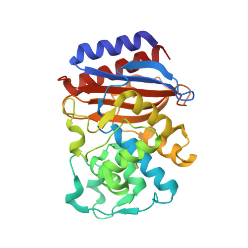
    5HVI, 5HW1, 5I52, 5I63, 5KKF, 5KPU, 6APA, 6AYK

  • PubMed Abstract: 

    Genetically encoded (GE) contrast agents detectable by magnetic resonance imaging (MRI) enable non-invasive visualization of gene expression and cell proliferation at virtually unlimited penetration depths. Using hyperpolarized 129 Xe in combination with chemical exchange saturation transfer, an MR contrast approach known as hyper-CEST, enables ultrasensitive protein detection and biomolecular imaging. GE MRI contrast agents developed to date include nanoscale proteinaceous gas vesicles as well as the monomeric bacterial proteins TEM-1 β-lactamase (bla) and maltose binding protein (MBP). To improve understanding of hyper-CEST NMR with proteins, structural and computational studies were performed to further characterize the Xe-bla interaction. X-ray crystallography validated the location of a high-occupancy Xe binding site predicted by MD simulations, and mutagenesis experiments confirmed this Xe site as the origin of the observed CEST contrast. Structural studies and MD simulations with representative bla mutants offered additional insight regarding the relationship between local protein structure and CEST contrast.


  • Organizational Affiliation

    Department of Chemistry, University of Pennsylvania, 231 S 34th St, Philadelphia, PA 19104.


Macromolecules
Find similar proteins by:  (by identity cutoff)  |  3D Structure
Entity ID: 1
MoleculeChains Sequence LengthOrganismDetailsImage
Beta-lactamase TEM
A, B, C, D
263Escherichia coliMutation(s): 1 
Gene Names: blablaT-3blaT-4blaT-5blaT-6
EC: 3.5.2.6
UniProt
Find proteins for P62593 (Escherichia coli)
Explore P62593 
Go to UniProtKB:  P62593
Entity Groups  
Sequence Clusters30% Identity50% Identity70% Identity90% Identity95% Identity100% Identity
UniProt GroupP62593
Sequence Annotations
Expand
  • Reference Sequence
Experimental Data & Validation

Experimental Data

  • Method: X-RAY DIFFRACTION
  • Resolution: 1.64 Å
  • R-Value Free: 0.206 
  • R-Value Work: 0.167 
  • R-Value Observed: 0.169 
  • Space Group: P 1 21 1
Unit Cell:
Length ( Å )Angle ( ˚ )
a = 60.663α = 90
b = 84.156β = 90.07
c = 95.703γ = 90
Software Package:
Software NamePurpose
PHENIXrefinement
XDSdata reduction
Aimlessdata scaling
PDB_EXTRACTdata extraction
PHASERphasing

Structure Validation

View Full Validation Report


Entry History & Funding Information

Deposition Data


Funding OrganizationLocationGrant Number
National Institutes of Health/National Institute of General Medical Sciences (NIH/NIGMS)United StatesR01-GM097478
Department of Defense Lung Cancer Research ProgramUnited StatesW81XWH-14-1-0424

Revision History  (Full details and data files)

  • Version 1.0: 2017-06-28
    Type: Initial release
  • Version 1.1: 2017-09-20
    Changes: Author supporting evidence
  • Version 1.2: 2019-01-16
    Changes: Data collection, Database references
  • Version 1.3: 2019-12-25
    Changes: Author supporting evidence
  • Version 1.4: 2023-09-27
    Changes: Data collection, Database references, Refinement description