5HM5

Crystal structure of Topo-97, an N-terminal 97kDa fragment of topoisomerase V


Experimental Data Snapshot

  • Method: X-RAY DIFFRACTION
  • Resolution: 2.40 Å
  • R-Value Free: 0.238 
  • R-Value Work: 0.199 
  • R-Value Observed: 0.201 

wwPDB Validation   3D Report Full Report


This is version 1.4 of the entry. See complete history


Literature

Methanopyrus kandleri topoisomerase V contains three distinct AP lyase active sites in addition to the topoisomerase active site.

Rajan, R.Osterman, A.Mondragon, A.

(2016) Nucleic Acids Res 44: 3464-3474

  • DOI: https://doi.org/10.1093/nar/gkw122
  • Primary Citation of Related Structures:  
    5HM5

  • PubMed Abstract: 

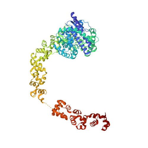
    Topoisomerase V (Topo-V) is the only topoisomerase with both topoisomerase and DNA repair activities. The topoisomerase activity is conferred by a small alpha-helical domain, whereas the AP lyase activity is found in a region formed by 12 tandem helix-hairpin-helix ((HhH)2) domains. Although it was known that Topo-V has multiple repair sites, only one had been mapped. Here, we show that Topo-V has three AP lyase sites. The atomic structure and Small Angle X-ray Scattering studies of a 97 kDa fragment spanning the topoisomerase and 10 (HhH)2 domains reveal that the (HhH)2 domains extend away from the topoisomerase domain. A combination of biochemical and structural observations allow the mapping of the second repair site to the junction of the 9th and 10th (HhH)2 domains. The second site is structurally similar to the first one and to the sites found in other AP lyases. The 3rd AP lyase site is located in the 12th (HhH)2 domain. The results show that Topo-V is an unusual protein: it is the only known protein with more than one (HhH)2 domain, the only known topoisomerase with dual activities and is also unique by having three AP lyase repair sites in the same polypeptide.


  • Organizational Affiliation

    Department of Molecular Biosciences, Northwestern University, 2205 Tech Drive, Evanston, IL 60208, USA.


Macromolecules
Find similar proteins by:  (by identity cutoff)  |  3D Structure
Entity ID: 1
MoleculeChains Sequence LengthOrganismDetailsImage
Topoisomerase V854Methanopyrus kandleriMutation(s): 0 
Gene Names: top5
UniProt
Find proteins for Q977W1 (Methanopyrus kandleri)
Explore Q977W1 
Go to UniProtKB:  Q977W1
Entity Groups  
Sequence Clusters30% Identity50% Identity70% Identity90% Identity95% Identity100% Identity
UniProt GroupQ977W1
Sequence Annotations
Expand
  • Reference Sequence
Experimental Data & Validation

Experimental Data

  • Method: X-RAY DIFFRACTION
  • Resolution: 2.40 Å
  • R-Value Free: 0.238 
  • R-Value Work: 0.199 
  • R-Value Observed: 0.201 
  • Space Group: P 1 21 1
Unit Cell:
Length ( Å )Angle ( ˚ )
a = 74.987α = 90
b = 77.3β = 93.09
c = 91.185γ = 90
Software Package:
Software NamePurpose
XDSdata reduction
SCALAdata scaling
SHARPphasing
PHENIXrefinement

Structure Validation

View Full Validation Report


Entry History & Funding Information

Deposition Data


Funding OrganizationLocationGrant Number
National Institutes of Health/National Institute of General Medical Sciences (NIH/NIGMS)United StatesR01GM51350

Revision History  (Full details and data files)

  • Version 1.0: 2016-03-09
    Type: Initial release
  • Version 1.1: 2016-05-04
    Changes: Database references
  • Version 1.2: 2017-09-13
    Changes: Author supporting evidence, Database references, Derived calculations
  • Version 1.3: 2019-12-25
    Changes: Author supporting evidence
  • Version 1.4: 2023-09-27
    Changes: Data collection, Database references, Refinement description