5HKG

Total chemical synthesis, refolding and crystallographic structure of a fully active immunophilin: calstabin 2 (FKBP12.6).


Experimental Data Snapshot

  • Method: X-RAY DIFFRACTION
  • Resolution: 1.50 Å
  • R-Value Free: 0.194 
  • R-Value Work: 0.172 
  • R-Value Observed: 0.173 

wwPDB Validation   3D Report Full Report


Ligand Structure Quality Assessment 


This is version 1.2 of the entry. See complete history


Literature

Total chemical synthesis, refolding, and crystallographic structure of fully active immunophilin calstabin 2 (FKBP12.6).

Bacchi, M.Jullian, M.Sirigu, S.Fould, B.Huet, T.Bruyand, L.Antoine, M.Vuillard, L.Ronga, L.Chavas, L.M.Nosjean, O.Ferry, G.Puget, K.Boutin, J.A.

(2016) Protein Sci 25: 2225-2242

  • DOI: https://doi.org/10.1002/pro.3051
  • Primary Citation of Related Structures:  
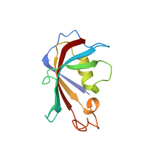
    5HKG

  • PubMed Abstract: 

    Synthetic biology (or chemical biology) is a growing field to which the chemical synthesis of proteins, particularly enzymes, makes a fundamental contribution. However, the chemical synthesis of catalytically active proteins (enzymes) remains poorly documented because it is difficult to obtain enough material for biochemical experiments. We chose calstabin, a 107-amino-acid proline isomerase, as a model. We synthesized the enzyme using the native chemical ligation approach and obtained several tens of milligrams. The polypeptide was refolded properly, and we characterized its biophysical properties, measured its catalytic activity, and then crystallized it in order to obtain its tridimensional structure after X-ray diffraction. The refolded enzyme was compared to the recombinant, wild-type enzyme. In addition, as a first step of validating the whole process, we incorporated exotic amino acids into the N-terminus. Surprisingly, none of the changes altered the catalytic activities of the corresponding mutants. Using this body of techniques, avenues are now open to further obtain enzymes modified with exotic amino acids in a way that is only barely accessible by molecular biology, obtaining detailed information on the structure-function relationship of enzymes reachable by complete chemical synthesis.


  • Organizational Affiliation

    Pôle d'Expertise Biotechnologie, Chimie and Biologie, Institut de Recherches Servier, 125 Chemin de Ronde, Croissy-sur-Seine, 78290, France.


Macromolecules
Find similar proteins by:  (by identity cutoff)  |  3D Structure
Entity ID: 1
MoleculeChains Sequence LengthOrganismDetailsImage
Peptidyl-prolyl cis-trans isomerase FKBP1B107Homo sapiensMutation(s): 0 
Gene Names: FKBP1BFKBP12.6FKBP1LFKBP9OTK4
EC: 5.2.1.8
UniProt & NIH Common Fund Data Resources
Find proteins for P68106 (Homo sapiens)
Explore P68106 
Go to UniProtKB:  P68106
PHAROS:  P68106
GTEx:  ENSG00000119782 
Entity Groups  
Sequence Clusters30% Identity50% Identity70% Identity90% Identity95% Identity100% Identity
UniProt GroupP68106
Sequence Annotations
Expand
  • Reference Sequence
Small Molecules
Ligands 3 Unique
IDChains Name / Formula / InChI Key2D Diagram3D Interactions
RAP
Query on RAP

Download Ideal Coordinates CCD File 
B [auth A]RAPAMYCIN IMMUNOSUPPRESSANT DRUG
C51 H79 N O13
QFJCIRLUMZQUOT-HPLJOQBZSA-N
EDO
Query on EDO

Download Ideal Coordinates CCD File 
F [auth A],
G [auth A],
H [auth A],
I [auth A]
1,2-ETHANEDIOL
C2 H6 O2
LYCAIKOWRPUZTN-UHFFFAOYSA-N
CL
Query on CL

Download Ideal Coordinates CCD File 
C [auth A],
D [auth A],
E [auth A]
CHLORIDE ION
Cl
VEXZGXHMUGYJMC-UHFFFAOYSA-M
Binding Affinity Annotations 
IDSourceBinding Affinity
RAP BindingDB:  5HKG Kd: 0.2 (nM) from 1 assay(s)
Experimental Data & Validation

Experimental Data

  • Method: X-RAY DIFFRACTION
  • Resolution: 1.50 Å
  • R-Value Free: 0.194 
  • R-Value Work: 0.172 
  • R-Value Observed: 0.173 
  • Space Group: P 21 21 21
Unit Cell:
Length ( Å )Angle ( ˚ )
a = 45.2α = 90
b = 48.61β = 90
c = 53.36γ = 90
Software Package:
Software NamePurpose
BUSTERrefinement
XDSdata reduction
SCALAdata scaling
MOLREPphasing

Structure Validation

View Full Validation Report


Ligand Structure Quality Assessment 


Entry History 

Deposition Data

Revision History  (Full details and data files)

  • Version 1.0: 2016-10-05
    Type: Initial release
  • Version 1.1: 2016-11-30
    Changes: Database references
  • Version 1.2: 2024-01-10
    Changes: Data collection, Database references, Refinement description