5HJQ

Crystal structure of the TBC domain of Skywalker/TBC1D24 from Drosophila melanogaster in complex with inositol(1,4,5)triphosphate


Experimental Data Snapshot

  • Method: X-RAY DIFFRACTION
  • Resolution: 2.30 Å
  • R-Value Free: 0.257 
  • R-Value Work: 0.230 
  • R-Value Observed: 0.231 

wwPDB Validation   3D Report Full Report


Ligand Structure Quality Assessment 


This is version 1.4 of the entry. See complete history


Literature

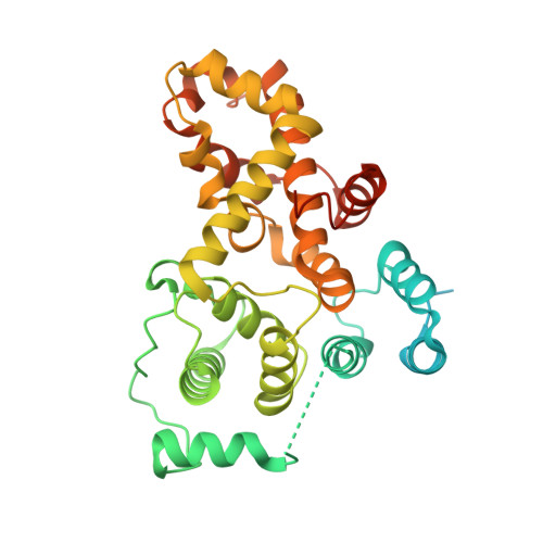
Skywalker-TBC1D24 has a lipid-binding pocket mutated in epilepsy and required for synaptic function.

Fischer, B.Luthy, K.Paesmans, J.De Koninck, C.Maes, I.Swerts, J.Kuenen, S.Uytterhoeven, V.Verstreken, P.Versees, W.

(2016) Nat Struct Mol Biol 23: 965-973

  • DOI: https://doi.org/10.1038/nsmb.3297
  • Primary Citation of Related Structures:  
    5HJN, 5HJQ

  • PubMed Abstract: 

    Mutations in TBC1D24 cause severe epilepsy and DOORS syndrome, but the molecular mechanisms underlying these pathologies are unresolved. We solved the crystal structure of the TBC domain of the Drosophila ortholog Skywalker, revealing an unanticipated cationic pocket conserved among TBC1D24 homologs. Cocrystallization and biochemistry showed that this pocket binds phosphoinositides phosphorylated at the 4 and 5 positions. The most prevalent patient mutations affect the phosphoinositide-binding pocket and inhibit lipid binding. Using in vivo photobleaching of Skywalker-GFP mutants, including pathogenic mutants, we showed that membrane binding via this pocket restricts Skywalker diffusion in presynaptic terminals. Additionally, the pathogenic mutations cause severe neurological defects in flies, including impaired synaptic-vesicle trafficking and seizures, and these defects are reversed by genetically increasing synaptic PI(4,5)P 2 concentrations through synaptojanin mutations. Hence, we discovered that a TBC domain affected by clinical mutations directly binds phosphoinositides through a cationic pocket and that phosphoinositide binding is critical for presynaptic function.


  • Organizational Affiliation

    Structural Biology Research Center, VIB, Brussels, Belgium.


Macromolecules
Find similar proteins by:  (by identity cutoff)  |  3D Structure
Entity ID: 1
MoleculeChains Sequence LengthOrganismDetailsImage
LD10117p376Drosophila melanogasterMutation(s): 0 
Gene Names: skyCG9339Dmel_CG9339
UniProt
Find proteins for Q9VIH7 (Drosophila melanogaster)
Explore Q9VIH7 
Go to UniProtKB:  Q9VIH7
Entity Groups  
Sequence Clusters30% Identity50% Identity70% Identity90% Identity95% Identity100% Identity
UniProt GroupQ9VIH7
Sequence Annotations
Expand
  • Reference Sequence
Small Molecules
Ligands 1 Unique
IDChains Name / Formula / InChI Key2D Diagram3D Interactions
I3P
Query on I3P

Download Ideal Coordinates CCD File 
B [auth A]D-MYO-INOSITOL-1,4,5-TRIPHOSPHATE
C6 H15 O15 P3
MMWCIQZXVOZEGG-XJTPDSDZSA-N
Binding Affinity Annotations 
IDSourceBinding Affinity
I3P Binding MOAD:  5HJQ Kd: 1.90e+4 (nM) from 1 assay(s)
Experimental Data & Validation

Experimental Data

  • Method: X-RAY DIFFRACTION
  • Resolution: 2.30 Å
  • R-Value Free: 0.257 
  • R-Value Work: 0.230 
  • R-Value Observed: 0.231 
  • Space Group: P 42 21 2
Unit Cell:
Length ( Å )Angle ( ˚ )
a = 87.72α = 90
b = 87.72β = 90
c = 97.097γ = 90
Software Package:
Software NamePurpose
PHENIXrefinement
XDSdata reduction
XDSdata scaling
PHASERphasing

Structure Validation

View Full Validation Report


Ligand Structure Quality Assessment 


Entry History 

Deposition Data

Revision History  (Full details and data files)

  • Version 1.0: 2016-09-21
    Type: Initial release
  • Version 1.1: 2016-10-05
    Changes: Database references
  • Version 1.2: 2016-11-16
    Changes: Database references
  • Version 1.3: 2018-01-24
    Changes: Advisory, Source and taxonomy
  • Version 1.4: 2024-01-10
    Changes: Advisory, Data collection, Database references, Refinement description