5HIW

Sorangium cellulosum So Ce56 cytochrome P450 260B1


Experimental Data Snapshot

  • Method: X-RAY DIFFRACTION
  • Resolution: 1.85 Å
  • R-Value Free: 0.219 
  • R-Value Work: 0.172 
  • R-Value Observed: 0.175 

wwPDB Validation   3D Report Full Report


Ligand Structure Quality Assessment 


This is version 1.1 of the entry. See complete history


Literature

Structure-function analysis for the hydroxylation of Delta 4 C21-steroids by the myxobacterial CYP260B1.

Salamanca-Pinzon, S.G.Khatri, Y.Carius, Y.Keller, L.Muller, R.Lancaster, C.R.Bernhardt, R.

(2016) FEBS Lett 590: 1838-1851

  • DOI: https://doi.org/10.1002/1873-3468.12217
  • Primary Citation of Related Structures:  
    5HIW

  • PubMed Abstract: 

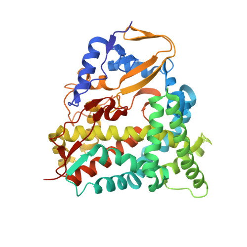
    Myxobacterial CYP260B1 from Sorangium cellulosum was heterologously expressed in Escherichia coli and purified. The in vitro conversion of a small focused substrate library comprised of Δ4 C21-steroids and steroidal drugs using surrogate bovine redox partners shows that CYP260B1 is a novel steroid hydroxylase. CYP260B1 performs the regio- and stereoselective hydroxylation of the glucocorticoid cortodoxone (RSS) to produce 6β-OH-RSS. The substrate-free crystal structure of CYP260B1 (PDB 5HIW) was resolved. Docking of the tested ligands into the crystal structure suggested that the C17 hydroxy moiety and the presence of either a keto or a hydroxy group at C11 determine the selectivity of hydroxylation.


  • Organizational Affiliation

    Institute of Biochemistry, Saarland University, Saarbrücken, Germany.


Macromolecules
Find similar proteins by:  (by identity cutoff)  |  3D Structure
Entity ID: 1
MoleculeChains Sequence LengthOrganismDetailsImage
Cytochrome P450 CYP260B1398Sorangium cellulosum So ce56Mutation(s): 0 
Gene Names: cypA3sce4806
EC: 1.14
UniProt
Find proteins for A9FFA1 (Sorangium cellulosum (strain So ce56))
Explore A9FFA1 
Go to UniProtKB:  A9FFA1
Entity Groups  
Sequence Clusters30% Identity50% Identity70% Identity90% Identity95% Identity100% Identity
UniProt GroupA9FFA1
Sequence Annotations
Expand
  • Reference Sequence
Small Molecules
Ligands 2 Unique
IDChains Name / Formula / InChI Key2D Diagram3D Interactions
HEM
Query on HEM

Download Ideal Coordinates CCD File 
B [auth A]PROTOPORPHYRIN IX CONTAINING FE
C34 H32 Fe N4 O4
KABFMIBPWCXCRK-RGGAHWMASA-L
MG
Query on MG

Download Ideal Coordinates CCD File 
C [auth A]MAGNESIUM ION
Mg
JLVVSXFLKOJNIY-UHFFFAOYSA-N
Experimental Data & Validation

Experimental Data

  • Method: X-RAY DIFFRACTION
  • Resolution: 1.85 Å
  • R-Value Free: 0.219 
  • R-Value Work: 0.172 
  • R-Value Observed: 0.175 
  • Space Group: P 31 2 1
Unit Cell:
Length ( Å )Angle ( ˚ )
a = 60.42α = 90
b = 60.42β = 90
c = 190.82γ = 120
Software Package:
Software NamePurpose
REFMACrefinement
iMOSFLMdata reduction
SCALAdata scaling
MOLREPphasing

Structure Validation

View Full Validation Report


Ligand Structure Quality Assessment 


Entry History 

Deposition Data

Revision History  (Full details and data files)

  • Version 1.0: 2016-08-03
    Type: Initial release
  • Version 1.1: 2024-01-10
    Changes: Data collection, Database references, Derived calculations, Refinement description