Distinct Features of Cyanophage-encoded T-type Phycobiliprotein Lyase Phi CpeT: THE ROLE OF AUXILIARY METABOLIC GENES.
Gasper, R., Schwach, J., Hartmann, J., Holtkamp, A., Wiethaus, J., Riedel, N., Hofmann, E., Frankenberg-Dinkel, N.(2017) J Biol Chem 292: 3089-3098
- PubMed: 28073912
- DOI: https://doi.org/10.1074/jbc.M116.769703
- Primary Citation of Related Structures:
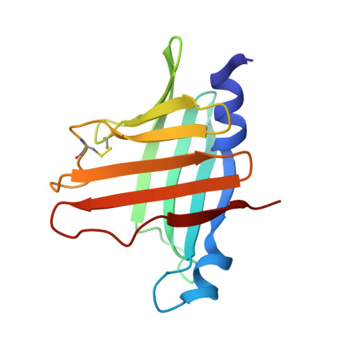
5HI8 - PubMed Abstract:
Auxiliary metabolic genes (AMG) are commonly found in the genomes of phages that infect cyanobacteria and increase the fitness of the cyanophage. AMGs are often homologs of host genes, and also typically related to photosynthesis. For example, the Φ cpeT gene in the cyanophage P-HM1 encodes a putative phycobiliprotein lyase related to cyanobacterial T-type lyases, which facilitate attachment of linear tetrapyrrole chromophores to Cys-155 of phycobiliprotein β-subunits, suggesting that ΦCpeT may also help assemble light-harvesting phycobiliproteins during infection. To investigate this possibility, we structurally and biochemically characterized recombinant ΦCpeT. The solved crystal structure of ΦCpeT at 1.8-Å resolution revealed that the protein adopts a similar fold as the cyanobacterial T-type lyase CpcT from Nostoc sp. PCC7120 but overall is more compact and smaller. ΦCpeT specifically binds phycoerythrobilin (PEB) in vitro leading to a tight complex that can also be formed in Escherichia coli when it is co-expressed with genes encoding PEB biosynthesis ( i.e. ho1 and pebS ). The formed ΦCpeT·PEB complex was very stable as the chromophore was not lost during chromatography and displayed a strong red fluorescence with a fluorescence quantum yield of Φ F = 0.3. This complex was not directly able to transfer PEB to the host phycobiliprotein β-subunit. However, it could assist the host lyase CpeS in its function by providing a pool of readily available PEB, a feature that might be important for fast phycobiliprotein assembly during phage infection.
Organizational Affiliation:
Protein Crystallography Group.